Computed Tomography (CT scan) types, uses, advantages and disadvantages
Computed tomography helps diagnose muscle and bone diseases, identifies the location of a tumor or clot, as well as detects and monitors diseases and injuries. The main types of scans are Skull tomography, Abdominal and pelvic tomography, Tomography of upper and lower limbs, and chest tomography. Computerized tomography (CT) scanning is similar to conventional radiology as it uses X-rays.
Computed Tomography
Computerized tomography (CT) is also referred to as computed tomography. A computed tomography scan is a medical imaging technique used to obtain detailed internal images of the body, The personnel that performs CT scans are called radiographers or radiology technologists.
Computerized axial tomography combines tomography imaging with computer techniques, The computed tomography (CT) scanner was invented by Godfrey Newbold Hounsfield in England, CT scan makes use of computer-processed combinations of many X-ray images taken from different angles to generate cross-sectional images.
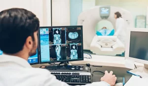
To obtain the computerized tomography scan, patients lie in the CT scanner – similar to a bed inside a ‘Polo® mint’, The X-ray tube and the detectors are opposite to each other, Both of these rotate around the patient, and information is obtained, usually, in slices.
The data are constructed by the computer and offer cross-sectional images in a single plane, which can be interpreted. The pictures are obtained by differences in X-ray absorption, and these differences are very small, allowing different shades of grey and distinction between different tissues.
Conventional radiography is a 2-dimensional image formed by the superimposition of images from successive layers of the body in the path of X-rays, The image of one layer is obscured by the superimposition of images of above and below layers, Tomography is invented to overcome this issue.
In the tomography technique, the images of selected layers are recorded sharply while images of other layers are recorded unsharply. The technique involves some form of movement of the patient or equipment during exposure, Tomography involves synchronized movement of any two of three subjects, viz. X-ray tube, film, and the patient while the third subject remains stationary.
Computed Tomography for CT scanning
CT scanner consists of an x-ray tube that emits a finely collimated fan-shaped x-ray beam directed through the patient to a series of scintillation detectors, These detectors measure the number of photons that exit the patient, The detectors form a continuous ring around the patient, and the X-ray tube moves in this circle within the fixed detector ring, The information is used to construct a cross-sectional image of the patient.
The radiation transmitted from the body of the patient is sensed by an array of detectors, The patient is made to move inside the chamber which contains an X-ray tube and detectors on a trolley, The detectors move in one axial direction, and the machine is called the computerized axial tomography (CAT).
The computer can reconstruct the images of the body layers from the output of the detectors. The above latest arrangement eliminates any need for the linear motion required to be given to the X-ray source and detectors which reduces scanning time drastically.
CT scanning uses X-rays to generate images of the body that are processed by a computer, which may be the bones, organs, or tissues, CT scanning does not cause pain, so it can be performed on anyone, Pregnant women should opt for other tests before computed tomography such as ultrasound or magnetic resonance imaging as radiation exposure is greater in CT.
Types of computerized tomography
There are two types of computerized tomography scans which are the Conventional CT scan and Spiral/helical CT scan, The Conventional CT scan can take slice by slice and after each slice, the scan stops and moves down to the next slice, such as from the top of the abdomen down to the pelvis, so, patients must hold their breath to avoid movement artifact. In a Spiral/helical CT scan, a continuous scan is taken in a spiral fashion, It is a much quicker process and the scanned images are contiguous.
Computerized tomography scans can be distinguished according to the plane in which the images are taken, computerised tomography scanning has been called ‘computed axial tomography (CAT)’ scanning, describing axial images that are taken – axial being the most common plane, other planes of imaging can be performed – eg, coronal or sagittal. The newer CT scanners can project these images into the 3D image.
Advantages of Computed Tomography
Computed Tomography eliminates the superimposition of images of undesired structures completely, CT offers higher contrast resolution, so, it can distinguish tissues having differences of less than 1% in their physical densities, Multiplanar reformatted imaging is possible due to multiple contiguous or single helical scans.
CT scanner offers more detail compared to ultrasonography, It is quicker, cheaper, and superior to MRI scanning, Motion artifacts are of less concern in CT scan than MRI. CT scanning helps diagnose various diseases, Physicians order CTs for various reasons, CT is made to scan the body part thought to be the source of the patient’s complaint or problem, in the hope that a diagnosis will somehow be reeled in.
Disadvantages of Computed Tomography
Computed Tomography is time-consuming, It is expensive for routine clinical use, and the patient is exposed to higher radiation, It requires expensive equipment and hence it is not always accessible at all levels of people, There is a risk of ionizing radiation and iodinated contrast agents.
Computerized tomography scanning requires breath holding which some patients can’t manage, Artefact is common – eg, metal clips, Computerised tomography scans of the brain can be affected by bone nearby, CT scanning is equivalent to 350 chest X-rays; CT abdomen to 400 chest X-rays and CT pulmonary angiography 750 chest X-rays. Children can suffer from leukaemia in mothers who have imaging during pregnancy.
The examination is done through the emission of radiation (X-rays) that can have harmful effects on health when the person is constantly exposed to this type of radiation. Radiologists and allied radiology personnel focused attention on the amount and potential risks of radiation that CT delivers.
CT is a costly and relatively high-dose procedure, with levels of radiation often approaching and sometimes exceeding those known to increase the probability of cancer, the radiation dose per procedure has not diminished with the advent of helical, fluoroscopic, and multi-slice techniques. While the evidence linking CT with cancer has not been established, the carcinogenic potential of this test is real.
How to prepare for the CT scan
Before performing the tomography, you should fast according to the doctor’s instructions, which may be from 4 to 6 hours so that there is better absorption of the contrast, You should stop the use of metformin if you are taking it, 24 hours before and 48 hours after the CT scan, since there may be a reaction with the contrast.
Computerized tomography scanning with contrast
Computed tomography uses contrast, which is a type of fluid that can be swallowed, injected into a vein, or inserted into the rectum during CT scanning to make it easier to see certain parts of the body. CT scanning offers images in shades of grey, but the shades are similar, making it difficult to discern between two areas, Contrast enhancement can be used to overcome this problem.
Side-effects of intravenous contrast
Injections are given rapidly and can cause a feeling of warmth in the arm, or even severe pain, Contrast can be extravasated, which can be severe enough to require skin grafting, Nausea, Vomiting, Urticaria, Anaphylaxis with bronchospasm, laryngeal oedema, hypotension, and Renal failure.
Contrast is excreted renally, If patients have chronic kidney disease, diabetes mellitus, or reduced intravascular volume then they run the risk of accumulating the contrast, This can lead to a worsening of renal impairment or even renal failure, good hydration before contrast will reduce the risk of developing renal impairment.
You can download Science online application on Google Play from this link: Science online Apps on Google Play
What is PET/CT, Positron emission tomography scan (PET) uses, risks and importance
Cone beam CT vs. Fan beam CT, CBCT advantages, disadvantages & uses
Interventional radiology types, Robotic endovascular systems advantages and disadvantages
Artificial intelligence in radiology decision support systems, Medical Imaging and healthcare
Artificial intelligence in medical field advantages & how AI medical diagnosis changes medicine
Applications of Artificial intelligence in the medical field and healthcare


