Visual pathway, functions of neurons in Primary visual cortex and Analysis of visual information
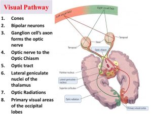
The visual nerve signals leave the retinae through the optic nerves. At the optic chiasma, the optic nerve fibers from the nasal halves of the retinae cross to opposite sides, where they join fibers from the opposite temporal retinae to form optic tracts.
Visual pathway
The fibers of each optic tract then synapse in the dorsal lateral geniculate body of the thalamus (LGN), and from there, geniculocalcarine fibers pass by the way of optic radiations (also called geniculocalcarine tract) to the primary visual cortex located mainly in the calcarine fissure area of the medial occipital lobe (Brodmann’s area 17), The areas surrounding it (areas 18 and 19 are the visual association areas.
Visual fibers give some collaterals that pass to other areas of the brain:
- The suprachiasmatic nucleus of the hypothalamus to control circadian rhythms that synchronize various physiologic changes of the body with night and day.
- To pretectal nuclei of the midbrain which is the center of the pupillary light reflex.
- To superior colliculus serving visual reflexes as eye and head movement and for control of rapid directional movements of the eyes.
The lateral geniculate body (LGB) of the thalamus
LGB is not only a relay station (3rd) order neuron in the visual pathway but also it receives cortical efferent fibers. This cortical projection to LGB may determine the type of visual stimuli that will be allowed to travel to consciousness and which are suppressed.
Primary visual cortex
Area 17 in the occipital lobe has an unusually thick layer IV because of large input from LGB. Layer IV is subdivided into a, b, c. Most of the projection from the thalamus goes to layer IV c. The visual cortex is organized into vertical columns of cells that detect lines at particular orientations in the visual field. Interspersed between these orientations columns are specific color blob areas for color vision.
The functions of neurons in the primary visual cortex V1
- Analysis of object color by blob neurons: neurons respond to a particular color or color differences (e.g. red versus green, yellow versus blue)
- Analysis of object shape or form: This is the function of simple and complex neurons that form the vertical columns of the primary visual cortex. This can detect lines, borders, edges, and angles that have specific orientation and position in the visual field. This is essential for form perception.
- Analysis of object motion: Some neurons respond best to a particular direction of motion, such as upward or downward, or leftward motion.
- Depth perception.
Secondary visual cortex
The primary visual cortex projects to area 18 and from there to other cortical association areas 21,20,19 and 7, These areas are essential for the analysis and interpretation of the retinal images.
The pathways for analysis of visual information
- Dorsal stream (position-gross form-motion pathway): After leaving the primary visual cortex, the signals flow into the posterior mid-temporal area and upwards to the occipitoparietal cortex, This pathway analyzes the third-dimensional position of the visual objects in space around the body, The pathway also analyzes a gross physical form of the visual scene as well as the motion of the scene.
- Ventral stream (analysis of visual details and colors): Impulses leaving the primary visual cortex pass to secondary visual areas of inferior, ventral, and medial regions of the occipital and temporal cortex, This is the pathway for the analysis of visual details, Separate areas dissect out color as well, The end result of information analysis in this pathway is to determine what the object is and what it means.
Visual field (Right and left visual hemifields)
The full visual field is the entire region of space that can be seen with both eyes looking straight ahead, fix your gaze on a point straight ahead, Now imagine a vertical line passing through the fixation point, dividing the visual field into left and right halves.
By definition, the objects appearing to the left of the midline are in the left visual hemifield, and objects appearing to the right of the midline are in the right visual hemifield. On looking straight ahead with both eyes open and then alternately closing one eye and then the other, you will see that the central portion of both visual hemifields is viewed by both retinas.
This region of space is therefore called the binocular visual field that can be divided into right and left hemifields, The objects in the binocular region of the left visual hemifield will be imaged on the nasal retina of the left eye and on the temporal retina of the right eye.
Because the fibers from the nasal portion of the left retina cross to the right side at the optic chiasma, all the information about the left visual hemifield is directed to the right side of the brain through the right optic tract. Each optic tract fibers represents the contralateral half of the visual field.
The optic nerve fibers cross in the optic chiasm so that the left visual hemifield is viewed by the right hemisphere and the right visual hemifield is viewed by the left hemisphere.
You can subscribe to Science Online on YouTube from this link: Science Online
You can download the Science Online application on Google Play from this link: Science Online Apps on Google Play
Retinal receptors, Photochemistry of vision, Steps of phototransduction in rods & Retinal output
Sense of vision, Refractive media of the eye, Corneal reflex & Pupillary pathways
Retina function, anatomy, layers, and accessory structures of the eye
Eyes structure, Histological organization of the fibrous and vascular coast of the eye
The visual system, Bony orbit anatomy, contents & Nerves, Muscles & movements of the Eyeball
Nystagmus types, causes, symptoms Development of CNS & Abnormalities of Spinal cord


