Urinary system structure, function, anatomy, organs, Blood supply and Importance of renal fascia
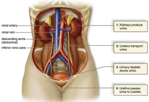
The urinary system consists of the kidneys, ureters, bladder, and urethra, It filters your blood, removing waste and excess water, and this waste becomes urine. the urinary system eliminates extra water and salt, toxins, and other waste products, The most common urinary issues are bladder infections and urinary tract infections (UTIs).
Urinary system
The urinary system is the system that is responsible for the secretion and excretion of excess water and waste products, outside the body in the form of urine.
Composition:
- 2 kidneys
- 2 ureters
- urinary bladder
- urethra
Kidney
It is the organ responsible for waste products, excess salt, and water from the blood.
Site of the kidney
Two kidneys lie in the upper posterior part of the abdominal cavity, one on each side. The right one lies lower than the left one by about 1-2 inches (2-5 cm) due to the presence of the liver in the right side which prevents its further ascent during its development.
Size: about the size of a clenched fist (length: 12 cm, width: 6 cm, thickness: 3 cm).
Shape: bean-shaped, having:
- 2 borders;
- The outer (lateral) border which is convex.
- The inner (medial) border which is convex up and down with a concavity in the middle (hilum).
- 2 poles (upper and lower).
- 2 surfaces (anterior and posterior).
Structures present in the Hilum
- The pelvis of the ureter (most posterior).
- Renal artery (in the middle).
- Renal vein (most anterior).
Surface anatomy of the kidneys
In the supine position: They extend from T12 to L3, the hilum lies at L1, The upper pole reaches the lower border of the 11th rib in the left side and the upper border of the 12th rib in the right side.
The rectangle of Morris is used to indicate the position of the kidney from the back; two vertical lines are drawn, the medial one lies 2.5 cm (1 inch) and the lateral one lies 9 cm (3.5 inches) from the midline; the parallelogram is completed by two horizontal lines drawn respectively at the levels of the tips of the spinous of the eleventh thoracic vertebra (T11) and the lower border of the spinous process of the third lumbar vertebra (L3), The hilum is 5 cm (2 inches) from the middle line at the level of the spinous process of the first lumbar vertebra (L1).
Relations of the kidneys
Anterior relations
Peritoneal covering
- Areas related to suprarenal glands and large intestine (both sides).
- Areas related to the duodenum (right kidney).
- Areas related to pancreas & splenic artery (left kidney).
Posterior relations
- Subcostal vessels
- Subcostal nerve.
- lliohypogastric nerve.
- llioinguinal nerve.
- Fibrous (true): It is intimately adherent to the kidney tissue.
- Fatty (perinephric fat): It is a fat surrounding the fibrous capsule.
- Fascial (Renal fascia): It surrounds the perinephric fat. It is a condensation of the areolar tissue between the peritoneum and the posterior abdominal wall and surrounding the perinephric fat.
- Paranephric fat: It is the fat surrounding the renal fascia. It is the extraperitoneal fat.
Clinical importance of renal fascia
- Sudden loss of weight may cause descent (ptosis) of the kidney due to a diminished amount of the perinephric fat.
- Pus spread does not occur downward as the anterior and posterior layers of the fascia join together to close the space inferiorly.
Blood supply of the kidney
- Arterial supply: by the renal artery that arises from the abdominal aorta at the level of L2.
- Venous drainage: through a renal vein that drains into the inferior vena cava.
- Kidney segmentation: it is a vascular segmentation of the kidney according to its arterial supply. There are five segments; apical, anterior superior, anterior inferior, posterior, and lower.
- Lymphatic drainage: To the para-aortic lymph nodes at the level of the L2 (origin of the renal artery).
You can subscribe to science online on YouTube from this link: Science Online
You can download Science Online application on Google Play from this link: Science Online Apps on Google Play
Suprarenal glands (adrenal glands) anatomy, structure & Effect of Deficiency of Catecholamines
Functions of Kidneys, Role of Kidney in glucose homeostasis, Lipid & protein metabolism
Histological structure of kidneys, Uriniferous tubules & Types of nephrons
Urine formation, Factors affecting Glomerular filtration rate, Tubular reabsorption & secretion
Urinary passages function, structure of Ureter, Urinary bladder & Uvulae vesicae


