Systemic Sclerosis types, causes, definition, diagnosis and treatment
Scleroderma is known as systemic sclerosis, It is a group of rare diseases that involve the hardening and tightening of the skin, It causes problems in the blood vessels, internal organs, and digestive tract, Scleroderma is categorized as “limited” or “diffuse,” which refers only to the degree of skin involvement, Both types can involve any of the other vascular or organ problems, Localized scleroderma, also known as morphea, affects only the skin.
Systemic Sclerosis
It is an auto-immune CT disorder (Complicated by Amyloidosis) characterized by: Degenerative and Inflammatory charges with subsequent intense Fibrosis (Fibrosis is the first which develops into sclerosis. It must be associated with: Skin with/without Vascular affection) affecting: Skin, Synovium, Blood vessels, skeletal muscles, GIT, Heart, Lung, and Kidney.
Etiopathogenesis
- Infections: Viral (CMV), Bacterial (Borellia that causes Lyme disease).
- Silica is an Industrial exposure. It also includes Gold, Coal, and Phenyl chloride (Used in tubes and wires).
- Epoxy resin is used in tires.
- Trichloroethylene is used in metal polishing.
- L-5-hydroxytryptophan (Used by athletes and old aged to build muscles: causes Eosinophilic Fasciitis (Fibrosis, Inflammation, and severe Pain in thighs with thick and Buckled skin, with fever, Myopathy and Neuropathy with no vascular affection (No Raynnauds or Pulmonary hypertension. In wedge biopsy for skin, underlying fascia and muscles: Eosinophile infiltration.
- Drugs: Chemotherapy (Bleomycin), Heroine addicts, Ergot and Tryptane (For migraine), and Beta blockers.
It is a genetic disease. Patients with HLA-DR52 have a bad prognosis, Genes are not enough to begin the disease, they must be associated with Environmental factors.
Fibroblasts include: Mesodermal, Endothelial, and Muscle cells. Inflammatory cytokine causes Chronic Inflammation. Collagen, proteoglycans, and fibronectin are secreted into: Skin layers and Walls of blood vessels.
Free radicals release cause: Chronic inflammation and Cancer. Vascular endothelial growth factor cell antibodies after deposition eventually leads to the narrowing of blood vessels and complete obstruction, causing Gangrene.
Criteria for classification (Diagnosis) of systemic sclerosis
The total score is determined by adding the maximum weight (score) in each category, Patients with a total score of ≥ 9 are classified as having definite scleroderma.
- If the patient scores 9 points or more: Definite Scleroderma.
- If the patient has the first criterion, it is enough to be diagnosed as Scleroderma.
- Pulmonary arterial hypertension may be either: Primary or Secondary (Due to interstitial pulmonary fibrosis) and it is the most common cause of death in Scleroderma.
- Anti-RNA polymerase 3 antibody is rare and associated with a bad prognosis.
Classification
Systemic sclerosis
A) With diffuse cutaneous scleroderma:
- Symmetric widespread thickening of the skin affecting Distal and Proximal extremities (Above Knees and Elbows), Trunk, Chest, Face, and Neck. There are areas of skin hyperpigmentation and hypopigmentation.
- Rapid progression of skin changes and system affection.
- Early appearance of visceral changes in lungs, kidneys, digestive system, and heart.
- Tendon friction rubs.
- Raynauds (75% of patients) at the same time or after the disease appearance.
- Diagnostic antibody: Antiscleroderma-70 (Anti-topoisomerase).
- Nail-fold capillary: Dilatation and capillary Destruction.
B) With Limited cutaneous scleroderma:
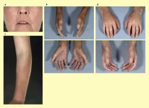
- Symmetric skin involvement is restricted to Distal extremities (Do not cross Elbows Knees) and Face (Face and Neck involvement are rare). Atrophic changes of the Ala nasi, Lips, Sacial amimia, and Telangiectasia of the skin.
- A slow progression of skin affection.
- Late appearance of visceral involvement. Late involvement of the lungs and late development of Pulmonary Hypertension.
- Raynaud’s (Vascular affection [Exagaeration] due to exposure to cold or stress). Universally: may preceed skin lesions by One year or more): It is characterized by three color changes of extremities (Fingers and Toes): White then Blue and pain due to ischemia that improves on warming (Raynauds + 2 positive specific antibodies [With no symptoms] = Limited Scleroderma).
- Dilated capillary loops in nail folds.
- Cutaneous calcification.
- Diagnostic antibody: Anti-centromere Antibody.
- Subtypes: Pulmonary Hypertension and Biliary Cirrhosis, and CREST:
- Cutaneous Calcinosis: Soft tissue deposition of calcium.
- Raynaud’s.
- Esophageal lesion.
- Sclerodactyly: Sclerodermal changes are limited to digits not extending beyond meta-carpophalangeal or Meta-tarsophalngeal joints (Fingers or Toes). Sausage-shaped and Loss of finger contour and non-pitting, due to deposition of collagen and proteoglycans in SC tissue not inflammation (Dactylitis is pitting: [Present in: Ankylosing spondylitis and Psoriasis]).
- Telangiectasia: Red spots on face, chest palm, and sole (All the body). It is not erythema. Macular dilatation of small blood vessels and capillaries. It becomes Blunched (Whiting) by pressure as it contains blood and by relieving pressure it returns red slowly due to slow fill with blood (Spider angioma Fills rapidly and returns red rapidly and is present in liver failure).
C) Sine scleroderma:
- Systemic manifestations with no skin lesions.
- It is a very rare type.
Localized scleroderma
Skin lesions without visceral lesions.
- Morphea: Plaque-like or Generalized. A third type is Gutted Morphen: Small circles of 1-2 mm. distributed on the upper chest and back shoulder, They are Waxy patches that are: Pale yellow or Brownish. It is Generalized: If it affects more than TWO sites of the body.
- Linear scleroderma. A line of fibrosis appears in the midline of the face (Hemi-atrophy of the face and the same side of the body).
Clinical Picture
- It has an insidious onset with worldwide distribution.
- Mainly Female. Female to Male (4:1).
- It is common in the fourth and fifth decades of age (40-50 years of age), More common in the 4th and 5th decades.
- Rarely visceral scleroderma occurs in absence of the skin changes (Sine Scleroderma).
Forms of Systemic Sclerosis
A) Limited Scleroderma:
- Skin thickening is distal to elbows and knees (To fingers and toes), not involving the trunk.
- Can involve perioral skin thickening (Pursing of lips).
- Less organ involvement.
- Isolated pulmonary hypertension (Pulmonary HTN) can occur.
- Seen in CREST syndrome: Calcinosis, Raynaud’s, Esophageal Dysmotility, Sclerodactyly, and Telangiectasia.
B) Diffuse Scleroderma:
- Skin thickening proximal to elbows and knees, involving the trunk.
- More likely to have organ involvement.
- Pulmonary fibrosis and Renal Crisis are more common.
Manifestations
Constitutional Manifestations:
It may be associated with;
- Mild fever.
- Anorexia.
- Weight loss.
- Insomnia: Due to pain (Skin, Ischemia and Musculoskeletal [Muscles and joints]).
- They cannot stretch their bodies due to skin stretching.
- Black and white spots all over the bodies (Hypo- and Hyper-Pigmentation): Salt and pepperpigmentation.
- Depression
Organ Involvement Manifestations
- Skin.
- Musculoskeletal.
- Pulmonary.
- Renal.
- Gastrointestinal.
- Cardiac.
- Gonadal system.
- Cancer.
- Vascular.
A-Skin Manifestations:
It appears in two stages:
Early-stage (Reversible: Anti-fibrotic agents and Immuno-suppressive agents) is characterized by itching and pitting edema of the skin due to water and salt retention.
Late stage (Irreversible) is characterized by no itching and non-pitting edema due to collagen, fibronectin, proteoglycans deposition and Salt & pepper pigmentation, telangiectasia, and digital ulcers. scars and loss of hair all over the body (Alopecia), loss of sweat (Fibrosis of sweat gland), and Calcinosis. Resorption of terminal phalanx (Due to continuous fibrosis and sclerosis of the finger and ischemia (Occurs also in leprosy, sarcoidosis, acromegaly, hyperparathyroidism) and loss of skin elasticity (Inability to make skin fold) and skin fixation to underlying structure (Skin tethering).
Pinched nose, fish mouth appearance, stretched checks, obliteration of naso-labial fold, Salt and pepper pigmentation, telangiectasia,
B-Musculoskeletal:
- Arthritis: in more than 50% of patients with Swelling, Morning stiffness, and Pain in the joints of the hands.
- Carpal Tunnel Syndrome: [Median nerve] Neuropathy in the lateral 3 and half fingers and part of the palm. The end stage is nerve lesion without regeneration and sensation loss.
- Contractures: Related to skin Thickening and Shortening of tendons and Tendinitis.
- Polymyositis: It may occur as part of mixed connective tissue disease or overlap.
- Normal joint structure: No swelling or erosion of articular cartilages no subchondral erosion (If not normal: Overlap with other disease: RA, gouty arthritis, psoriatic arthritis, septic arthritis).
- Tendon rub due to tendinitis (Hard grating sound): It occurs in active, progressive and bad prognostic patients and Patients with systemic affection and overlap with RA.
- Proximal myopathy: Muscle weakness proximal to digitals and thighs.
C-Pulmonary
- Pneumonitis due to food regurge from the esophagus.
- It is the leading cause of death since we are better at controlling renal disease.
- Symptoms: Exertional dyspnea.
- Cancer Lang.
- Pulmonary embolism.
- Types of lung Involvement: Basal Interstitial lung disease (In alveoli [fibrosing alveoli] and base of the lung), and Isolated pulmonary hypertension (First cause of death): Either primary or Secondary.
D-Renal Manifestations
- Scleroderma Renal Hypertensive Crisis (Second cause of death after pulmonary hypertension).
- Sudden acute oliguric (Anuric) renal failure.
- Abruptly developing severe hypertension [Acute pulmonary edema, cerebrovascular stroke, encephalopathy, severe headache and migraine, blurring of vision, and epileptic episodes]: It is treated by Blood dialysis and Renal transplantation: Rise in SBP by (>30 mmHg) and DBP by (> 20 mm Hg).
- It is characterized by an increase in serum creatinine by 50% over baseline or creatinine > 120 of the upper limit. Proteinuria more than 2+ by dipstick. Hematuria more than 2+ by dipstick or more than 10 RBC/HPF. Thrombocytopenia less than 100 [Micro-vasculopathy due to clot formation], Hemolysis (Schisetocytes, Low platelets, Increased Reticulocyte count), RBCs casts, CBC will show Shisctocytes [fragmented RBCs] (Micro-angiopathic hemolytic anemia) that block renal blood vessels.
- Can cause Headache, Encephalopathy, Seizures, and Left ventricular failure.
- 90% of patients with blood pressure over 150/90.
- Can also occur with lower blood pressure or normohypertension: Less than 140/90 and this confers a worse prognosis.
- Risk Factors for Renal Crisis: They require regular BP measurement. This patient must take ACE inhibitors or APR as a protective measure.
- Rapidly progressive skin thickening within the first 2-3 years.
- Steroid use (prednisone > 15 mg).
- Anti-polymerase III Ab.
- Pericardial Effusion.
E- Gastrointestinal
- Disordered peristalsis of the lower two-thirds (Dysphagia) of the esophagus presents as dysphagia (Due to fibrosis and sclerosis), leading to chocking, dry cough, retrosternal chest pain and harshness of voice.
- Impaired function of the lower esophageal sphincter (Fibrosis and sclerosis), leading to: food regurge and erosion of esophageal mucosa and esophageal ulcers (PRE-CANCER) and microaspiration [Food reaches the base of the lung].
- Chronic esophageal reflux includes erosive chronic esophagitis with bleeding. Barrett’s esophagus) and lower esophageal stricture.
- Involvement of the stomach occurs in systemic sclerosis and presents clinically as ease of satiety and on occasion as either functional gastric outlet obstruction or acute gastric dilatation. Also presented by difficulty of digestion and rupture of gastric arteries (Chronic Hematemsis causing iron deficiency anemia)
- Ectezia (GAVE: gastric antrum vascular ectezia) of gastric vessels in the fundus of the stomach: tortuous, dilated, congested, and ruptured causing hematemis. It is diagnosed before rupture by upper endoscopy and appears as a watermelon appearance of the stomach.
The most common types of cancer in this disease are cancer skin, Cancer esophagus, Cancer colon, and Cancer lung.
Small bowel involvement
- Intermittent bloating abdominal cramps, intermittent or chronic diarrhea, and presentations suggestive of intestinal obstruction.
- Malabsorption occurs.
- Bacterial overgrowth (Feeds on Vitamin B12 and Folic acid, causing Pernicious anemia) in areas of intestinal stasis occurs frequently (In intestinal loops).
Large bowel involvement
- Constipated stools and eventually diarrhea.
- Intra colonic pressure increases causing diverticulosis and diverticulitis [Pre cancer] (Form pockets into anterior abdominal wall muscles).
F. Cardiac Manifestations
- Pericardial Effusion (Bad prognosis: active patient and rapidly progressive, leading to Renal hypertensive crisis) and Symptomatic constrictive pericarditis in 20% of patients.
- Micro-vascular Coronary Artery Disease: Recurrent vasospasm of coronary arteries, Necrosis, and Patchy myocardial fibrosis leads to: Diastolic more than Systolic dysfunction.
- Myocarditis: Inflammation which leads to fibrosis.
- Ventricular Arrhythmias, pre-mature pace, Ventricular fibrillation. Ventricular tachycardias and conduction abnormalities (Heart block), due to: Fibrosis of the cardiac conduction system and AV conduction defects and arrhythmias.
- Endocarditis.
- Restrictive cardiomyopathy with systolic and diastolic dysfunction of the heart.
- Scleroderma is an un-independent risk for coronary artery disease.
G. Vascular affection
Most affected: Peripheral [Raynnauds, Ischemia, and Gangrene], Coronary, Pulmonary, Renal, Gastric and Digital.
H-Gonadal
They are completely fertile. Males have Erectile Dysfunctions (EDs) due to muscle fibrosis and vascular affection of the penis. Females have painful intercourse due to: vaginal fibrosis and dry vaginal secretion penis.
Investigations
Laboratory:
- Increased ESR (Rare or Mild).
- CBC: 5 types of anaemia: (Iron deficiency, Auto-immune, Traumatic, Micro-angiopathic [Most fatal and danger]). Thrombocytopenia.
- Increased Creatine Phosphokinase levels in patients with muscle involvement.
- Increased Urea and Creatinine levels in patients with kidney involvement.
- Serology:
- Anti-centromere (Limited type).
- Anti-scleroderma 70 [Anti-topoisomerase) (Diffuse type).
- Anti-nuclear (Diffuse type).
- Anti PM [Polymyositis]- Scleroderma (Overlap).
- Hypo-complementaemia and Anti-ds-DNA: RARE (Overlap).
- Positive RF (Overlap).
- Increased Gamma Globulin.
- Anti-RNA polymerase [Any type is Bad prognostic].
Synovial fluid
- Increased WBCs (Less than 10,000).
- Increased plasma cells.
- Increased lymphocytes.
- Fibrin deposits.
- Decreased mucin.
- Decreased viscosity.
Wide field nail fold capillary microscope
- Loss of capillaries, Dilatation and Tortusity.
- Capillary microscopy: Enlarged capillaries are observed in all 3 portions of the capillary nail fold: Arterial, Apical and Venous, especially at the Edge of the nail fold. Adjacent areas are avascular.
Bone resorption
Bone radiography reveals generalized osteopenia, which most commonly affects the hands Intra-articular calcifications are observed.
You can subscribe to Science Online on YouTube from this link: Science Online
You can download the Science Online application on Google Play from this link: Science Online Apps on Google Play
Systemic Lupus Erythematosus symptoms, diagnosis, types and treatment
Rheumatoid Arthritis (RA) causes, stages, symptoms, diagnosis, and specific joint affection
Rheumatoid Arthritis diagnosis and treatment, Disease-Modifying antirheumatic drugs (DMARDs)
Non-radiographic axial spondyloarthritis, Reactive arthritis, and Enteropathic arthritis


