Synaptic transmission steps, Synapses types and Nature of the postsynaptic change
Communications between neurons in the central nervous system occur through synapses. A synapse is a specialized functional junction between two neurons. In the nervous system, there are two types of synapses: electrical and chemical synapses.
Electrical synapse
They are extremely rare in the nervous system. At an electrical synapse, the membranes of the presynaptic and postsynaptic neurons come close together forming areas of fusion called gap junctions. In these junctions, the opposing membranes are held in position by connecting protein channels called connexons, which contain pores that permit ions to pass directly from one cell to the other. This allows rapid direct conduction of electrical potential from one neuron to the next as if no membrane barrier exists.
Chemical synapse
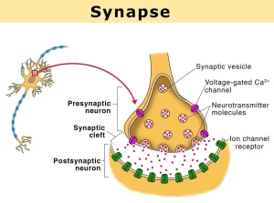
Almost all synapses in the human central nervous system are chemical synapses. In these synapses, the impulses in the presynaptic axon cause secretion of a chemical transmitter at the synapse, and this transmitter act on receptors located in the membrane of the postsynaptic neuron to modify its activity either exciting or inhibiting it depending upon the nature of the postsynaptic receptors.
Mechanism of neurotransmitter release from a synapse
When an action potential arrives at the synaptic knob, depolarization occurs, which increases the permeability of the knob to Ca2+ through the opening of voltage-gated Ca2+ channels leading to the influx of Ca2+ ions into the synapse. This causes the release of the neurotransmitter by exocytosis proportional to the Ca2+ influx.
The neurotransmitter diffuses through the synaptic cleft and binds to receptor molecules on the postsynaptic membrane leading to the opening of chemical-gated channels for specifications that are allowed to pass the membrane causing a small local change in the membrane potential of the postsynaptic neuron.
Nature of the postsynaptic change
During rest, the postsynaptic neuron is in the polarized state, showing resting membrane potential (RMP) of about 70-mv. The threshold for its excitation is about 59-mv. The neurotransmitter released at the synapse will lead to either:
- Excitation of the postsynaptic neuron if the released transmitter is excitatory e.g. acetylcholine, noradrenaline, and serotonin.
- Inhibition of the postsynaptic neuron if the released transmitter is inhibitory e.g. gamma-aminobutyric acid (GABA) and glycine.
Electrical events in excitatory synapses
The binding of the excitatory transmitter to postsynaptic receptors increases the permeability of the postsynaptic membrane to all ions, but the movement of Na+ predominates due to its large electrochemical gradient across the membrane.
The Na+ influx results in slight depolarization of the postsynaptic neuron, which will increase its excitability and bring it closer to the threshold. This change in voltage above RMP is called excitatory postsynaptic potential (EPSP).
Electrical events in inhibitory synapses
1. Postsynaptic inhibition
The binding of the inhibitory transmitter to postsynaptic receptors increases the permeability of the postsynaptic membrane to K+ and CL−. The resulting K+ outflux and CL− influx increases the degree of intracellular negativity leading to a state of hyperpolarization at the postsynaptic neuron causing its inhibition. This hyperpolarization state is called the inhibitory postsynaptic potential (IPSP).
2. Presynaptic inhibition
The inhibitory response may occur in the presynaptic terminals before the neuronal signals reach the postsynaptic neuron. This process is mediated by special interneurons, which terminate on excitatory presynaptic terminals of other neurons forming axo-axonic synapses with these terminals before they end on the postsynaptic neurons.
Most of the neurons which inhibit the presynaptic terminals release the inhibitory neurotransmitter gamma amino butyric acid which inhibits the release of the excitatory transmitter from the excitatory presynaptic fiber and thereby decreasing the postsynaptic response.
Central excitatory and inhibitory state
Most postsynaptic neurons are covered with thousands of synaptic knobs derived from a large number of presynaptic neurons. Some of these presynaptic inputs are excitatory while others are inhibitory to the postsynaptic neuron. If a neuron, at a given instant, receives more excitatory impulses than the inhibitory, there will be a central excitatory state, in which the neuron is depolarized. On the other hand, if the neuron receives more inhibitory impulses than the excitatory, there will be a central inhibitory state, in which the neuron is hyperpolarized.
Factors affecting synaptic transmission
a. Effect of pH
Alkalosis markedly increases neuronal excitability and synaptic transmission e.g. in persons predisposed to epileptic attacks, hyperventilation (which washes out CO2 and raises blood pH) leads to convulsions. Acidosis greatly depresses neuronal excitability and synaptic transmission e.g. acidosis that occurs in diabetic and uremic patients often leads to coma.
b. Effect of hypoxia
Hypoxia for only a few seconds can result in complete in-excitability of neurons. Loss of consciousness occurs if the cerebral circulation is temporarily interrupted for 3-6 seconds.
c. Effect of drugs
Some drugs increase the excitability of neurons and enhance synaptic transmission e.g. caffeine, theophylline, and theobromine (found in coffee, tea, and cocoa) which act by decreasing the threshold for excitation of neurons. Other drugs decrease neuronal excitability and synaptic transmission e.g. hypnotics and anesthetic drugs. decreasing the postsynaptic response.
Features of synaptic transmission, Mechanism of sensitization & long term potentiation
Physiology of the cerebral cortex, Wernicke’s area, Broca’s area, sensory & motor areas
Histological structure of Cerebral cortex & Types of neurons in the cerebral cortex
Blood supply of CNS (central nervous system), Carotid system & Circle of Willis
Referred pain examples, Causes of neuropathic pain & Pain control mechanisms
Physiology of central human reflexes, Types & properties of Spinal cord reflexes
You can follow science online on YouTube from this link: Science online
You can download Science Online application on Google Play from this link: Science Online Apps on Google Play


