Suprarenal (adrenal) cortex hormones, structure, function and location
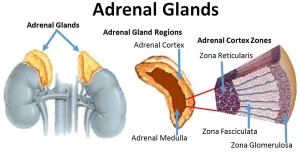
The adrenal gland releases certain hormones directly into the bloodstream, It is made of two main parts: The adrenal cortex & the adrenal medulla, The adrenal cortex is the outer region, it is the largest part of the adrenal gland, It consists of three separate zones: zona glomerulosa, zona fasciculata, and zona reticularis, Each zone is responsible for producing specific hormones, The adrenal medulla produces “stress hormones,” including adrenaline.
Suprarenal (adrenal) cortex
It is the steroid-secreting portion of the suprarenal gland. Histologically, It is divided into 3 zones:
A- Zona glomerulosa
By LM
- It lies immediately under the connective tissue capsule, forming 15% of the cortex
- The cells are arranged in closely packed arched clusters surrounded by fenestrated blood capillaries.
- The cells are columnar or pyramidal in shape with dense nuclei and slightly vacuolated eosinophilic cytoplasm.
By EM: the cells show the ultrastructural features of steroid-secreting cells i.e. sER, mitochondria with tubular cristae, and few lipid droplets. Function: the cells secrete mineralocorticoids, mainly aldosterone.
B. Zona fasciculata
By LM:
- It is the middle and the largest layer of the cortex, forming 65% – 80 % of the cortex.
- The cells are arranged into long straight cords, perpendicular to the capsule.
- The cords are one or two cells thick and are separated longitudinally. arranged blood sinusoidal capillaries.
- The cells are large, polyhedral, with large, pale spherical nuclei.
- The cytoplasm is pale eosinophilic and contains large amount of lipid droplets, which appear as numerous vacuoles and so these cells are called spongiocytes).
By EM: the cells show the ultrastructural features of steroid secreting cells i.e. abundant sER, mitochondria with tubular cristae, and numerous lipid droplets. Function: It secretes glucocorticoids.
C- Zona reticularis
By LM
- It is the inner layer of the cortex, forming 7% of the cortex.
- Its cells are arranged in irregular short cords that anastomose together enclosing fenestrated capillaries in between.
- The cells are small with eosinophilic cytoplasms containing few vacuoles.
- Dark cells are seen near the suprarenal medulla. They have dark cytoplasm with pyknotic nuclei, suggesting that they are degenerating cells.
By EM: the cells show the ultrastructural features of steroid-secreting cells with few lipid droplets. Lipofuscin pigments are characteristic in this layer.
Function: It secretes androgens.
Mineralocorticoid (Aldosterone)
Aldosterone is a steroid. It is secreted from the zona glomerulosa. It is the most important hormone that regulates Na+ and K+ metabolism, and it combines loosely with the plasma proteins.
Functions of the mineralocorticoid (Aldosterone)
1. On Kidney
It increases Na+ concentration and decreases K+ concentration in the extracellular fluid by acting on the kidney. Aldosterone increases sodium reabsorption in exchange with the secretion of either K+ or H+ from the distal tubules, collecting tubules and collecting duct. It increases the formation of Na+ K+ ATPase.
2. On sweat glands, salivary glands, and intestinal cells.
Aldosterone increases the reabsorption of sodium and the secretion of potassium by the ducts, The effect on the sweat glands is important to conserve body salt in hot environments and the effect on the salivary glands is necessary to conserve salt when excessive quantities of saliva are lost, Aldosterone also greatly enhances sodium absorption by the intestines, which obviously prevents the loss of sodium in the stools.
Regulation of Aldosterone secretion
The four factors in the regulation of aldosterone secretion are:
- Potassium ion concentration of the extracellular fluid.
- Renin-angiotensin system.
- Quantity of body sodium.
- Adrenocorticotropic hormone (ACTH).
I. Effect of potassium ion concentration on aldosterone secretion: An increase in potassium ion concentration causes direct stimulation of zona glomerulosa to increase secretion of aldosterone, which increases excretion of potassium, therefore, the potassium ion concentration returns to its normal level.
II. Effect of the renin-angiotensin system on aldosterone secretion: Angiotensin II is responsible for stimulating the synthesis and release of aldosterone from the cells of the zona glomerulosa. The rate of aldosterone secretion is effectively determined by the rate of secretion of renin from the juxtaglomerular cells.
III. Effect of decreased sodium on aldosterone secretion:
- Diminished sodium concentration directly affects the zona glomerulosa cells to enhance the aldosterone secretion.
- Lack of sodium causes retention of potassium by the kidneys which increases aldosterone secretion.
- Diminished sodium leads to diminished extracellular fluid volume, with resultant diminished cardiac output and renal blood flow, This enhances the formation of angiotensin II which stimulates aldosterone secretion.
- Diminished sodium concentration causes the anterior pituitary gland to secrete some substances called the unidentified pituitary factor which affects the adrenal glands to increase aldosterone secretion.
IV. Effect of ACTH on aldosterone secretion: It has a permissive effect on aldosterone secretion and all the above regulations of aldosterone secretion will occur if there is only a minimal amount of ACTH.
Effects of hypersecretion of aldosterone (hyperaldosteronism)
- Decrease in sodium loss in the urine with increase sodium in extracellular fluid, Excess aldosterone causes increase sodium reabsorption, this also causes reabsorption of chloride. Excess sodium chloride causes increased osmotic pressure so osmosis of water occurs. Absorption of sodium and water increases extracellular fluid volume and blood volume which may lead to increase cardiac output and hypertension.
- Increase potassium loss in the urine: The excessive loss of potassium ions from the extracellular fluid into the urine causes a serious decrease in the plasma potassium concentration called hypokalemia, When the potassium ion concentration falls, severe muscle weakness often develops. Paralysis may occur due to the failure of transmission of the action potential.
- Excess aldosterone causes tubular secretion of hydrogen ions instead of sodium causing a decrease in hydrogen ion concentration in extracellular fud which leads to a mild degree of alkalosis, The condition is known as Conn’s syndrome.
Effects of hyposecretion of aldosterone (hypoaldosteronism)
- Decrease potassium loss in the urine: There is a failure of excretion of potassium ions instead of sodium. Potassium ions concentration can rise far above normal (called hyperkalemia). When it rises to approximately double normal, serious cardiac toxicity including weakness of heart contraction and development of arrhythmia and if severe cardiac arrest occurs in diastole.
- Mild acidosis develops due to excess hydrogen ions in the blood due to a decrease in its excretion instead of sodium.
- Increase in sodium loss in the urine with decrease sodium in extracellular fluid. When aldosterone is deficient, sodium reabsorption decreases so sodium, chloride ions, and water are lost in the urine. This leads to:
- Decreased extracellular fluid volume.
- Decreased plasma volume.
- Decreased cardiac output.
- Circulatory shock may develop rapidly.
You can follow science online on YouTube from this link: Science online
Glucocorticoid (cortisol) function, Effects of cortisol on skeletal muscle and circulatory system
Suprarenal glands (adrenal glands) anatomy, structure and Effect of Deficiency of Catecholamines
Calcium metabolism, Calcitonin function, Vitamin D3 and Biosynthesis of calcitriol
Parathyroid gland anatomy, structure, function, hormones and Tetany types
Histological structure of thyroid gland and Functions of Thyroid Hormones (T3 & T4)
Endocrine system structure, function, disorders, Endocrine Glands and Hormone types


