Stretch reflex (myotatic reflex) types, properties, Muscle tone, muscle spindles and Clonus
Reflexes, or reflex actions, are involuntary, almost instantaneous movements in response to a specific stimulus, They protect your body from things that can harm it. The reflex action is also known as a reflex, When a person accidentally touches a hot object, they automatically jerk their hand away without thinking, The reflex does not require any thought input.
Stretch reflex (myotatic reflex)
It is the reflex contraction of the skeletal muscle in response to passive stretch; i.e. when the skeletal muscle with an intact nerve supply is stretched, it contracts.
Innervation of the muscle spindle
Sensory innervation (afferent fibers): two kinds of sensory axons innervate the central receptor area of the muscle spindle:
- Primary or annulospiral fibers: they encircle the central receptor area of the nuclear bag and nuclear chain. They are peripheral termination of the myelinated la sensory fibers, which are the most rapidly conducting nerve fibers in the entire body.
- Secondary or flower spray ending: these fibers arise from the peripheral part of the receptor area of the nuclear chain fibers. They are the termination of the smaller type II myelinated sensory fibers.
Motor innervation (efferent fibers): the motor innervation of the intrafusal fibers is supplied by gamma efferent fibers that arise from small motor neurons in the ventral horn of the spinal cord [gamma motor neurons (yMNs)]. These neurons innervate the peripheral contractile regions of the intrafusal fibers. There are two types of gamma-efferent fibers:
- Dynamic gamma efferent: they supply the nuclear bag fibers.
- Static gamma efferent: they supply the nuclear chain fibers.
Alpha motor neurons: these are the center of the stretch reflex. They receive input from the primary and secondary endings when the spindles are stretched and discharge their impulses along thick myelinated fibers to the extrafusal muscle fibers.
Mechanism of stimulation of the muscle spindles
The adequate stimulus that excites the muscle spindles is the stretching of their central parts. This can occur in 2 ways:
- Stretching of the whole muscle.
- Activation of the gamma efferent fibers, which causes contraction of the peripheral parts of the intrafusal fibers leading to stretch of the receptor area.
Importance of γ motor innervation
Stimulation of γ-motor neurons produces a very different picture from that produced by stimulation of the a-motor neurons: Stimulation of y-motor neurons does not lead directly to detectable contraction of the muscles because the intrafusal fibers are not strong enough or plentiful enough to cause shortening, However, stimulation does cause the contractile ends of the intrafusal fibers to shorten and therefore stretches the nuclear bag portion of the spindles, deforming the endings, and initiating impulses in the la fibers.
This in turn can lead to reflex contraction of the muscle, Thus, muscles can be made to contract via stimulation of the α-motor neurons that innervate the extrafusal fibers or the γ-motor neurons that initiate contraction indirectly via the stretch reflex.
Increased y-motor neuron activity thus increases spindle sensitivity during the stretch. In response to descending excitatory input to spinal motor circuits, both α- and γ-motor neurons are activated, Because of this “α-γ coactivation” intrafusal and extrafusal fibers shorten together (servo-assistant device), and spindle afferent activity can occur throughout the period of muscle contraction.
Properties of the stretch reflex
- Its total reflex time is short because it is monosynaptic and its afferent and efferent fibers are rapidly conducting.
- It is localized i.e. shows no irradiation due to the absence of interneurons.
- It shows reciprocal innervations.
- Shows no after discharge i.e. the muscle contraction stops immediately on releasing the stretching force due to the absence of interneurons.
- Shows no fatigue because during stretch the motor neurons discharge at a low frequency and alternate their activity.
Types of stretch reflex
A. Static stretch reflex (Muscle tone)
It is a neurogenic, maintained, subtetanic skeletal muscle contraction during rest. Tone is present in all skeletal muscles of the body, but it is more marked in the antigravity muscles being additionally stretched by the effect of gravity. These muscles are: the flexors of the upper limbs, the extensors of the lower limbs, the extensors of the back and neck, and elevators of the lower jaw.
Based on the static response of the secondary endings to stretch: This reflex occurs throughout the period of steady stretch and is produced mainly as a result of the stretch of the nuclear chain fibers (slowly adapting receptors). The response is an increase in the rate of discharge from the secondary endings and continues as long as the stretch is maintained. It provides information about the steady length of muscle fibers.
Functions of the muscle tone
- It maintains an erect posture against the pulling effect of gravity.
- It helps both venous return and lymph flow from the lower parts of the body, (muscle pump).
- It helps heat production, so it increases in cold weather (shivering) and decreases in hot weather.
- The abdominal muscle tone keeps the viscera in position.
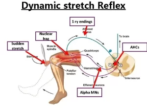
B. Dynamic stretch reflex (Tendon jerk)
This includes the deep reflexes for example the knee jerk and ankle jerk. It is one of the motor tests used to diagnose and localize neurological lesions.
Based on the dynamic response of the primary endings to stretch: This reflex occurs while the muscle length is actually increasing, and is produced mainly as a result of the stretch of the nuclear bag fibers (rapidly adapting receptors). The response is a rapid increase in the rate of discharge from the primary endings as they are very sensitive to the velocity of the change in muscle length during a stretch followed by a marked decrease when the new length is maintained. It provides information about the speed of movements to allow for quick corrective movements.
Static stretch reflex
- Stimulus: Weak maintained passive stretch as the distance between the muscle origin and its insertion is longer than its actual length.
- Receptor: Nuclear chain.
- Afferent: Flower spray ending
- Center: Alpha Motor Neuron of Anterior Horn of Spinal Cord.
- Efferent: Thick my elinated axons of alpha motor neuron.
- Effecter: Extrafusal muscle fibers.
- Response: Muscle tone which is neurogenic, maintained. subtetanic skeletal muscle contraction.
Dynamic stretch reflex
- Stimulus: Sudden strong passive stretch.
- Receptor: Nuclear bag.
- Afferent: Annulospiral ending.
- Center: Alpha Motor Neuron of Anterior Horn of Spinal Cord.
- Efferent: Thick my elinated axons of alpha motor neuron.
- Effecter: Extrafusal muscle fibers.
- Response: Tendon jerks which are rapid contraction followed by rapid relaxation.
Higher (supraspinal) control of the stretch reflex
Facilitatory supraspinal discharge:
- Medial reticulospinal which activates the gamma motor neurons
- The vestibular nuclei activate the alpha motor neurons
- Efferent connection from the cerebellum to the vestibular nuclei may modulate muscle stretch reflex and tone.
- Motor area 4 activates the alpha motor neurons.
Inhibitory supraspinal discharge:
- The lateral medullary reticular formation which inactivates the gamma motor neurons.
- Premotor area.
- Efferent connections from the cerebellum to the reticular formation may inhibit muscle stretch reflex and tone.
The stretch reflex is regulated by many supraspinal facilitatory and inhibitory centers which discharge downwards through the descending tracts to affect the activity of the motor neurons, Through such higher control, the tone of various muscles can be changed, which is important for keeping the posture of the body.
Other factors also influence γ-motor neuron discharge
Anxiety causes an increased discharge, a fact that probably explains the hyperactive tendon reflexes sometimes seen in anxious patients. It is well known that trying to pull the hands apart when the flexed fingers are hooked together facilitates the knee-jerk reflex (Jendrassik’s maneuver) and this may be due to increased γ-motor neuron discharge initiated by afferent impulses from the hands.
Inverse stretch reflex (Golgi tendon reflex)
Whereas the stretch reflex causes muscle contraction in response to increased muscle length (stretch), Golgi tendon reflexes produce exactly the opposite effect, muscle relaxation and lengthening in response to increased muscle tension. An increase in muscle tension leads to excitation of the high threshold Golgi tendon organs (GTOs), tension receptors in the tendon, which fire impulses along thick myelinated afferent (1b).
The lb fibers end in the spinal cord on inhibitory interneurons that in turn terminate directly on alpha motor neurons inhibiting them, As a result, the contracting muscle relaxes, It is an example of a disynaptic reflex. The Golgi tendon reflex is a protective reflex that helps to avoid the tearing of muscles and tendons subjected to possibly damaging stretching force.
Clonus
It is rhythmic alternative muscle contraction and relaxation, It develops in response to sudden maintained stretch of spastic paralyzed muscle due to increased γ-motor neuron discharge.
Mechanism of clonus
Clonus is the result of a stretch reflex inverse stretch reflex sequence, The initial sudden muscle stretch will stimulate discharge from intrafusal fibers (mainly nuclear bag) resulting in muscle contraction (stretch reflex).
The tension developed in the muscle as a result of contraction is so high that the Golgi tendon organ becomes excited initiating the inverse stretch reflex and producing relaxation, As the stretch is maintained, this process will be repeated rhythmically helped by the state of excessive supraspinal facilitation in case of upper motor neuron lesion.
Reflex actions importance
Reflex actions are incredibly important for our survival. They are involuntary responses to stimuli that happen very quickly, without having to think about them. This is because the reflex arc, the pathway that signals travel through during a reflex, bypasses the brain and instead goes through the spinal cord.
Reflex actions are an essential part of our nervous system. They help to keep us safe, healthy, and functioning properly. Reflex actions help us to react quickly and safely in dangerous situations. Some examples of reflex actions are coughing, Sneezing, Gagging, Swallowing, Pupil dilation, and constriction
Some reasons why reflex actions are important:
Speed: Reflexes are much faster than voluntary actions, This is because they bypass the brain and instead travel through the spinal cord. This allows us to react to danger much more quickly than we could if we had to think about what to do first. if you touch a hot stove, you will pull your hand away before you consciously realize that it’s hot. This split-second difference can be enough to prevent serious injury.
The blink reflex, which protects your eye from getting injured, takes only about 1/10 of a second. If you had to think about blinking every time something came near your eye, you would likely get injured much more often.
Protection: Many reflexes are designed to protect us from harm. the knee-jerk reflex helps to keep us balanced and prevent our legs from buckling when we trip, and the blink reflex protects our eyes from dust and debris, the withdrawal reflex causes you to pull your hand away from a hot stove before you feel the heat. This helps to prevent burns and other injuries.
Homeostasis: Reflexes also help to maintain homeostasis, or a stable internal environment, which is the body’s ability to keep its internal conditions stable. the knee-jerk reflex helps to keep us upright and balanced, the pupil reflex helps to regulate the amount of light that enters the eye, and the gag reflex helps to prevent us from choking on food or other objects.
You can follow Science online on YouTube from this link: Science online
You can download Science online application on Google Play from this link: Science online Apps on Google Play
Physiology of central human reflexes, Types & properties of Spinal cord reflexes
Physiology & function of the spinal cord, Lateral & medial brainstem pathway
Histological organization spinal cord, Relation between spinal & vertebral segments
Features of synaptic transmission, Mechanism of sensitization & long term potentiation
Synaptic transmission steps, Synapses types & Nature of the postsynaptic change
Blood supply of CNS (central nervous system), Carotid system & Circle of Willis
Anatomy of brainstem, Features of medulla oblongata, pons & midbrain


