Stones in the urethra, Grades of Renal Injury, and Ureteral stone treatment without surgery
Stones in the urethra, also known as urethral calculi, are uncommon. They are more likely to occur in men than in women due to the narrower urethra in males. These stones can form anywhere along the urethra, but they are most commonly found near the opening at the tip of the penis.
Urethral stones
Stones in the urethra originate in the kidneys or the bladder and then travel down the urinary tract, They can also form in the urethra itself due to infection or a foreign object. Treatment for urethral stones involves removing the stone. This may be done in a variety of ways, depending on the size and location of the stone.
In some cases, the stone may be small enough to pass on its own with increased fluids. For larger stones, medications to relax the urethra or procedures to break up the stone with shock waves (lithotripsy) may be needed.
Pathogenesis and Clinical Presentation
- A stone that enters the urethra from the bladder is always small. It either passes out with urine during voiding or is impacted at any level in the urethra.
- An impacted stone causes obstructive urinary symptoms.
Diagnosis:
1. Palpation of the stone:
- Stone in the penile urethra: is felt along the ventral surface of the shaft of the penis.
- Stone in the bulbous urethra: is felt in the perineum) (behind the scrotum).
- Stone in the prostatic urethra: may be felt by digital rectal examination.
2. A plain X-ray of the urethra: shows a radio-opaque stone.
3. (Ascending retrograde) urethrogram: shows a radiolucent stone as a filling defect.
Treatment
- A stone in the prostatic urethra is pushed back during endoscopy and is then treated as a small bladder stone by endoscopic stone crushing.
- Surgical removal of a urethral stone is allowed only if the endoscopy failed.
- A stone in the anterior urethra near the external meatus is extracted after performing a meatotomy.
Injuries to the urinary tract
Injuries to the kidney
The kidney is the most common organ injured in the urinary tract. Most renal traumas are managed conservatively.
Etiology:
- Blunt injury (80%) following a road traffic accident (RTA) or falling from a height.
- Penetrating injury (20%) from a gunshot bullet or a stab wound by a knife.
- latrogenic, e.g.: during endoscopy or renal biopsy.
Grades of Renal Injury: (done after CT abdomen & pelvis with contrast)
- Grade 1: contusion or contained sub-capsular hematoma without laceration
- Grade II: perinephric hematoma+/- cortical laceration less than 1 cm, по urine extravasation.
- Grade III: cortical laceration more than 1 cm without any urine extravasation.
- Grade IV: urine extravasation with a cortical laceration or thrombosis segmental vessel.
- Grade V: shattered kidney, or renal pedicle avulsion or thrombosis, or pelvi-ureteric junction avulsion.
Grades I, II, III (85%) of cases)…minor renal injuries usually managed conservatively. Grades IV, V (15% of cases)… major renal injuries and usually require surgical intervention.
Clinical Presentation
- Loin pain from collection of a hematoma with or without urinoma.
- Total hematuria if the injury involves a calyx or the renal pelvis. The intensity of hematuria does not correlate with the severity of renal injury. Usually absent in grade I, II, III and in cases with renal artery injury or pelviureteric avulsion.
- Abdominal swelling, distention, rigid abdomen.
- Retroperitoneal hematoma can be assessed by palpation and if it is pulsatile and increasing in size this denotes progressive internal hemorrhage and mandates immediate exploration.
- The patient may present in shock.
Complications
- Uncontrolled hemorrhage with compromised vital signs.
- Urinoma.
- Perinephric abscess.
- Late complication: reno-vascular hypertension from increased (renin secretion secondary to infarction of lacerated devascularized renal tissue, hydronephrosis, renal atrophy.
Diagnosis
- C.T. scan with contrast for the abdomen and pelvis is the most reliable tool for the diagnosis of renal injury. It can detect: the extent of renal injury, size and extent of retroperitoneal hematoma, vascular injury, an infarcted renal segment, associated injury of other abdominal organs, and status of the other kidney.
- IVU, ultrasound, and renal angiography can detect the above findings if done combined.
- Laboratory investigations: urine analysis, serial Hematocrit, serum creatinine, and blood urea.
Treatment
1. Grades I-II-II: conservative treatment is recommended and includes bedrest, antibiotics, and maintenance of vital signs. Usually, the injury resolves spontaneously, and no intervention is needed.
2. Grade IV:
- If the patient is vitally stable conservative treatment is indicated with close follow-up.
- If the patient is vitally unstable immediate surgical intervention is indicated to suture the lacerated segment if possible otherwise nephrectomy is done.
3. Grade V: it’s a life-threatening condition and immediate surgical exploration with nephrectomy is mandatory. In the case of avulsed PUJ alone (repair over a DJ is done, if not possible PCN is inserted.
4. Delayed surgical treatment of complications:
- If hypertension occurs from an infarcted renal segment. The infarcted segment is surgically removed.
- Persistent urinoma: ureteric double J stent insertion and drainage.
- The peri-renal abscess must be drained.
- PUJ stenosis is treated surgically (endopyelotomy)
- Secondary hydronephrosis is treated by PCN.
Injuries to the ureter
Parts of the ureter
The ureter is composed of three parts:
- Upper 1/3 extends from the pelviureteric junction till the lower border of the fourth lumbar vertebra.
- Middle 1/3 extends from the lower border of the fourth lumbar vertebra to the lower border of the sacroiliac joint.
- Lower 1/3 extends from the lower border of the sacroiliac joint till the bladder in an oblique course.
Etiology:
Ureteric trauma may be:
1. External Trauma: (Usually affects the upper and middle thirds).
- Blunt trauma e.g. road traffic accident or falling from a height.
- Sharp trauma e.g. stab wound or bullet.
II. Internal trauma: (Usually affects the lower third)
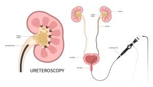
- Endoscopic e.g. during ureteroscopy.
- Abdominal surgery e.g. GIT, gynecological or obstetric surgeries.
Grades of ureteric injury
- Contusion in the wall.
- Partial injury.
- Complete ureteric cut.
Presentation
- History of the cause.
- Hematuria (microscopic or gross).
- Flank pain on the affected side is usually due to urinary collection.
- Anuria if bilateral injury occurred.
- Intraoperative: urine in the operative field, look for the rent in the ureteric wall and look for a suture occluding the ureter.
- During ureteroscopy: Mucosal injury or ureteric wall perforation.
- Delayed causes may present with ureteric stricture and hydronephrosis urinoma and abscess formation, urinary fistula loss of kidney function.
Investigations
- Laboratory investigations including urine analysis, blood urea, and serum creatinine.
- CT with contrast is the most reliable diagnostic tool to assess the level of ureteric injury, detect the site and extent of urine leakage, and assess the amount of urinoma.
- Ultrasonography can detect urinoma collection.
- IVU is used to detect dye extravasation from the ureter.
- An ascending ureterogram will detect the escape of the contrast at the site of perforation.
Treatment
Factors influencing the treatment decision:
- The time of detecting the injury.
- The level of the ureteric injury.
- The grade of ureteric injury.
For small partial ureteric injuries that are early detected at any level insertion of a DJ is indicated. Immediate recognized large ureteral trauma:
Surgical exploration with debridement, tension-free and watertigh ureteric anastomosis
- Lower third injuries: ureteric reimplantation in the bladder.
- Middle-third injuries: repair of the ureter over a ureteric stent (direct anastomosis between the cut ends),
- Upper third ureter: repair of the ureter over a ureteric stent.
Delayed recognition of the trauma 7-10 days:
- Drainage of the urinoma.
- Urinary diversion: percutaneous nephrostomy.
Definitive Delayed repair usually after 3-6 months:
- Ureteric replacement by intestinal segment in case of long segment injury.
- Transureteroureterosotomy (anastomosing the injured ureter with the normal ureter on the other side).
- Nephrectomy is indicated when the renal function is lost due to long-standing stricture formation.
You can subscribe to Science Online on YouTube from this link: Science Online
You can download Science Online application on Google Play from this link: Science Online Apps on Google Play
Urolithiasis, Stones in the kidney or ureter, How do you treat urolithiasis?
Anomalies of the bladder and urethra, Urinary tract abnormalities symptoms
What are urinary tract infections?, kidney problems, Urinary retention and Bladder stones
Functions of Kidneys, Role of Kidney in glucose homeostasis, Lipid & protein metabolism
Histological structure of kidneys, Uriniferous tubules and Types of nephrons
Urine formation, Factors affecting Glomerular filtration rate, Tubular reabsorption and secretion
Urinary passages function, structure of Ureter, Urinary bladder & Uvulae vesicae
Urinary system structure, function, anatomy, organs, Blood supply and Importance of renal fascia
Urinary bladder structure, function, Control of micturition by Brain & Voluntary micturition


