Salivary glands function, shape, lobes, surfaces and Structures within the parotid gland
Saliva has an enzyme called amylase that makes it easier for the stomach to break down starches in food. It helps moisten food so we can swallow it more easily. Saliva prevents infections in the mouth and throat. It has an important role in our oral health, It helps to maintain healthy teeth and prevent bad breath, It moistens the oral cavity, which helps with swallowing and speaking.
Salivary glands
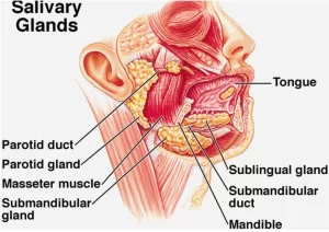
The paired parotid glands, together with the paired submandibular and sublingual glands and the numerous small glands scattered throughout the mouth cavity, constitute salivary glands.
Parotid gland
The parotid gland is the largest gland of the salivary glands, there is a small part that lies over the parotid duct is called the accessory part of the gland.
Shape and lobes and surfaces of the gland
The parotid gland is wedge-shaped, with its base above and its apex behind the angle of the mandible. It has three surfaces:
- Superficial surface.
- Anteromedial surface.
- Posteromedial surface.
Boundaries:
- Upwards: Zygomatic arch.
- Downwards: Angle of the mandible (just below it).
- Anterior: Masseter (overlies its posterior part).
- Posterior: Sternomastoid (overlies its upper part).
Capsules of the gland:
The gland is enclosed in a dense fibrous capsule derived from the investing layer of deep cervical fascia. The investing deep cervical fascia splits at the angle of the mandible into: a superficial layer and a deep layer which is the stylomandibular ligament. This separates the parotid gland from the submandibular gland.
Applied anatomy:
That is why a swelling in the parotid gland becomes very painful because it is enclosed between the splitted two layers of deep fascia.
Parotid duct:
The parotid duct emerges from the anterior border of the gland and passes forward over the lateral surface of the masseter muscle, At the anterior border of the muscle, it turns sharply medially and pierces the buccal pad of fat and buccinator muscle, It then opens into the vestibule of the mouth opposite the upper second molar tooth.
Surface anatomy of parotid duct
The parotid duct corresponds to the middle third of a line extending between two points:
- A point midway between the red margin of the upper lip and ala of the nose.
- A point at the lower end of the tragus of the ear.
Structures within the parotid gland
1- Facial nerve: (the most superficial structure)
The facial nerve enters the gland through the posteromedial surface, Then, it breaks to form a plexus (pes anserinus) inside the parotid gland, The plexus gives a group of terminal branches which leave the gland through:
- Upper end: Temporal branches.
- Anterior border. Zygomatic, buccal, and, mandibular branches.
- Lower end: Cervical branch.
2- Retromandibular vein intermediate in position
The superficial temporal vein enters the parotid gland through the upper end, The maxillary vein enters the gland through the antero-medial surface, The two veins form the retromandibular vein inside the gland, and the retro-mandibular vein leaves the gland through the lower end as the anterior and posterior branches.
3- External carotid artery (the deepest structure)
The external carotid artery enters the gland through the postero-medial surface, It divides inside the gland into the maxillary and superficial temporal arteries, the superficial temporal artery leaves the gland through the upper end, the maxillary artery leaves the gland through the antero-medial surface.
4- Auriculotemporal nerve
It enters the gland through the antero-medial surface and leaves it through the upper pole.
5- Parotid L.N.
Blood supply: The external carotid artery and the terminal branches within the gland, namely, superficial temporal and maxillary arteries, supply the gland, and the veins drain into the retromandibular vein.
Lymph drainage: The lymph vessels drain into the parotid lymph nodes and the deep cervical lymph nodes.
Nerve supply:
Parasympathetic: the fibres pass through the glossopharyngeal nerve, They leave the nerve in its tympanic branch; which reach the tympanic cavity where it breaks into the tympanic plexus. The fibers leave the plexus through lesser superficial petrosal nerve which leaves to the infratemporal fossa through the foramen oval then relay in the otic ganglion. Postganglionic fibers reach the gland through the auriculotemporal nerve.
Sympathetic fibers: reach the gland as a plexus of nerves around the external carotid artery.
Sensory:
- For the capsule ———— Great auricular nerve.
- For parenchyma ———— Auriculo-temporal nerve.
Relations of the parotid gland
- Superficial (lateral) relations are skin, superficial fascia containing platysma, parotid lymph nodes and great auricular nerve.
- Posteromedial relations are the mastoid process, sternocleidomastoid, and posterior belly of the digastric. More medially styloid process and its attached three muscles. carotid sheath containing the internal carotid artery, internal jugular vein and last cranial nerves and facial nerve trunk.
- Anteromedial relations are the posterior border of the ramus of the mandible, masseter, and medial pterygoid muscle. The parotid duct leaves through this surface.
Submandibular gland
The submandibular gland is a large salivary gland that lies partly under the cover of the body of the mandible and it is made up of a large superficial part and a small deep part, which are continuous with each other around the posterior border of the mylohyoid muscle.
I. The superficial part of the gland
It lies in the digastric triangle. Posteriorly, it is separated from the parotid gland by a stylomandibular ligament.
Relations of the superficial part of the gland
- Superficial or inferolateral surface: Skin, superficial fascia containing platysma, and the investing layer of the deep cervical fascia, cervical branch of the facial nerve, Submandibular lymph nodes and anterior facial vein.
- Medial surface: it is related medially to mylohyoid muscle, mylohyoid nerve and vessels, hyoglossus muscle, lingual and hypoglossal nerves and submandibular ganglion.
- Lateral surface: it lies in contact with the submandibular fossa on the medial surface of the mandible, facial artery/which lies between the gland and the mandible, and the medial pterygoid.
II. The deep part of the gland
Relations of the deep part of the gland
It extends forwards between mylohyoid and hyoglossus muscles below the lingual nerve and above the hypoglossal nerve.
Submandibular duct
The submandibular duct arises from the medial surface of the superficial part of the gland, Near the posterior border of the mylohyoid, it runs with the deep part of the gland, It passes forward between the mylohyoid and hyoglossus along the side of the tongue.
Beneath the mucous membrane of the floor of the mouth, It is crossed laterally by the lingual nerve then below then medial to the duct (triple relation) and then lies between the sublingual gland laterally and genioglossus muscle medially, It opens into the floor of the mouth on the summit of a small sublingual papilla, which is situated at the side of the frenulum of the tongue.
Blood supply
It is supplied by branches of the facial and lingual arteries, The veins drain into the facial and lingual veins.
Lymph drainage
Lymph vessels drain into the submandibular and the deep cervical lymph nodes.
Nerve supply
Parasympathetic secretomotor supply passes through the seventh cranial nerve (facial nerve) via the chorda tympani nerve with Relay in the submandibular ganglion and Postganglionic fibers go to submandibular and sublingual salivary glands.
Sublingual salivary gland
The sublingual gland is the smallest of the three main salivary glands, It lies beneath the mucous membrane of the floor of the mouth, close to the midline.
- Sublingual ducts: The sublingual ducts open into the mouth on the summit of the sublingual fold.
- Blood supply: The gland is supplied by branches of the facial and lingual arteries, The veins drain into the facial and lingual veins.
- Lymph drainage: Lymph vessels drain into the submandibular and the deep cervical lymph nodes.
- Nerve supply: Similar to submandibular Parasympathetic gland.
Postganglionic fibres pass through the lingual nerve.
You can download Science online application on Google Play from this link: Science Online Apps on Google Play
Git function, Types of salivary secretion & Composition of saliva
Histological structure of salivary glands, Parotids, Sublingual and Submandibular glands
Norma lateralis features & anatomy, Mandible surfaces, nerves & ligaments
Mouth Cavity divisions, anatomy, function, muscles, Contents of Soft palate and Hard palate
Tongue function, anatomy & structure, Types of lingual papillae & Types of cells in taste bud


