Posterior abdominal wall muscles, layers, blood supply and anatomy
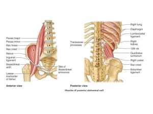
The posterior abdominal wall is formed by the lumbar vertebrae, pelvic girdle, posterior abdominal muscles, and their associated fascia, Significant vessels, nerves, and organs are located on the inner surface of the posterior abdominal wall. There are five muscles in the posterior abdominal wall: the iliacus, psoas major, psoas minor, quadratus lumborum, and diaphragm.
Posterior abdominal wall
Muscles of the posterior abdominal wall:
Muscle: PsoasMajor
- Origin: Transverse processes of a lumbar vertebra; the lateral surface of bodies of T12-L5 and intervening IV discs.
- Insertion: By a strong tendon in the lesser trochanter of the femur.
- Innervation: Lumbar plexus via the anterior rami of (L1- 2- 3) nerves.
- Actions: Flexion of the thigh, Lateral flexion of the trunk.
Muscle: PsoasMinor (May be absent)
- Origin: Lateral surface of bodies of T12-L1 and intervening IV discs.
- Insertion: Pectineal line and ilio-pectineal eminence.
- Innervation: Anterior rami of L1.
- Actions: Weak flexion of the trunk.
Muscle: lliacus
- Origin: Upper two-thirds of iliac fossa, ala of sacrum, anterior sacroiliac, and iliolumbar ligaments.
- Insertion: Lesser trochanter of femur and shaft inferior to it. (With the tendon of the psoas major muscle).
- Innervation: Femoral nerve (L2-L4).
- Actions: Flexes thigh and stabilizes the hip joint, acts with the psoas major.
Muscle: Quadratus lumborum
- Origin: lliolumbar ligament, the internal lip of the iliac crest, and transverse processes of L5.
- Insertion: Medial half of inferior border of 12th rib and tips of lumbar transverse processes.
- Innervation: Anterior rami of T12 and L1 L4 nerves.
- Actions: Laterally flexes vertebral column and fixes 12th rib during inspiration.
Abdominal aorta
The abdominal aorta is the largest artery in the abdominal cavity, It is a direct continuation of descending thoracic aorta, It is 13 cm long, It enters the abdomen opposite the 12th thoracic vertebra through the aortic opening of the diaphragm, It ends by dividing into 2 common iliac arteries opposite the 4th lumbar vertebra.
Relations of the abdominal aorta: Anterior relations from superior to inferior:
- Coeliac ganglia and plexus.
- Body of the pancreas.
- Splenic and left renal veins.
- (3rd part) part of the duodenum.
- Superior mesenteric vessels and root of the mesentery.
Posterior relations:
- Lumbar vertebrae (1-4) and intervertebral discs.
- Anterior longitudinal ligament.
- 3rd and 4th lumbar veins.
Branches of abdominal aorta
- Inferior phrenic arteries, L1 (upper border), Paired.
- Coeliac trunk, L1 (upper border), Single.
- Superior mesenteric artery, L1 (lower border), Single.
- Middle suprarenal arteries, L1 (lower border), Paired.
- Renal arteries, L2, Paired.
- Gonadal arteries, L3 lower level of L2 upper level of I3, Paired.
- Four lumbar arteries, L1-L4, Paired.
- Inferior mesenteric artery, L3, Single.
- Median sacral artery, L4, Single.
- Common iliac arteries, L4, Paired.
The formation of the inferior vena cava is at L5 (below that of the bifurcation of the aorta).
Common Iliac Arteries
The abdominal aorta divides, on the left side of the body of the fourth lumbar vertebra, into the two common iliac arteries, Each artery ends in front of the sacroiliac joint by dividing into external and internal iliac arteries.
Inferior vena cava (IVC)
- It is the largest vein in the body, It is formed by the union of two common iliac veins anterior to and just to the right of the 5th lumber vertebra.
- It ascends on the right side of the aorta, passes through the caval opening of the diaphragm opposite T8, and drains into the right atrium.
- It conveys blood from the whole body below the diaphragm to the right atrium.
Tributaries of I.V.C
- Two common iliac veins.
- Two pairs of lumbar veins: 3rd, 4th.
- Two renal veins (Rt. & Lt.).
- Two inferior phrenic veins.
- Two hepatic veins.
- Right gonadal vein.
- Right suprarenal vein.
- Median sacral vein.
Nerves of the posterior abdominal wall
Several important components of the nervous system are in the posterior abdominal region, These include the sympathetic trunks and associated splanchnic nerves, the plexus of nerves and ganglia associated with the abdominal aorta, and the lumber plexus of nerves.
Sympathetic Trunk
- Two long ganglionated nerve strands one on each side of the vertebral column.
- It extends from the base of the skull to the coccyx.
- It enters the abdomen deep to the medial arcuate ligament of the diaphragm.
- It lies along the medial border of psoas major. Anteriorly on the right side, it lies behind the I.V.C. and on the left side, it lies along the left side of the aorta.
- It passes inferiorly behind common iliac vessels.
- Its termination is by joining to form the unpaired ganglion impar (anterior to coccyx).
Lumbar plexus
♦ Formation: formed by anterior rami of L1-L3, and upper part of L4.
♦ Position: lies within the substance of psoas major, Therefore, relative to the psoas major muscle the various branches emerge either:
- Anterior-genitofemoral nerve;
- Medial- obturator nerve and lumbosacral trunk.
- Lateral-iliohypogastric, ilioinguinal, femoral nerve, and lateral cutaneous nerve of the thigh.
The iliohypogastric nerve
The iliohypogastric nerve is formed by fibers from L1. It supplies the skin over the lateral gluteal region and the skin above the pubis.
The ilioinguinal nerve
The ilioinguinal nerve is formed in common with the iliohypogastric nerve (L1). It passes through the superficial inguinal ring.
The genitofemoral nerve
The genitofemoral nerve is formed from L1, 2 and passes through the psoas major to emerge on its anterior surface, It runs downwards on the psoas and divides into genital and femoral branches, The genital branch enters the inguinal canal through the deep inguinal ring to supply the cremasteric muscle and a small area of overlying skin, The femoral branch passes behind the inguinal ligament to enter the femoral sheath and supply the skin over the femoral triangle.
Lateral cutaneous nerve of the thigh
It is formed from the posterior division of the L2, 3. The lateral cutaneous nerve of the thigh emerges at the lateral border of the psoas muscle. It supplies the skin on the lateral part of the thigh.
Femoral nerve
The femoral nerve arises from the posterior division of the L2, 3, 4 anterior primary rami, The nerve lies in the groove between the psoas and iliacus and enters the thigh behind the inguinal ligament.
Obturator nerve
The nerve arises from the anterior division of the L2, 3, and 4 anterior primary rami, It emerges medial to the psoas muscle and leaves the pelvis through the obturator foramen.
Lumbosacral trunk
A part of the anterior division of L4 and L5 primary rami forms the lumbosacral trunk, The trunk lies anterior to the ala of the sacrum to join S1 anterior primary ramus. (sacral plexus).
Muscular nerves
T12 and lumbar primary rami send short nerves into neighboring muscles; quadratus lumborum, and psoas.
You can download Science online application on google Play from this link: Science online Apps on Google Play
Regulation of stomach emptying, Causes of vomiting & Nervous pathway of reflex vomiting
Constituents of the gastric juice, Gastric motility & types of movements occur in the stomach
Physiology & functions of Stomach, Composition of gastric secretion
Histological structure of stomach, Fundic glands of Stomach & Gastric musculosa
Abdomen muscles, Blood Supply of Anterior Abdominal Wall & Rectus Sheath content
Stomach parts, function, curvatures, orifices, peritoneal connections & Venous drainage of Stomach


