Physiology and functions of Cerebellum, Cerebellar lesions, Motor and Non-motor functions
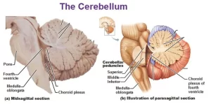
The cerebellum plays a role in regulating the planning and execution of movement. It receives inputs from various areas of the body as well as the cortex and projects to the brainstem and motor areas of the cortex via the thalamus The cerebellar motor functions are executed automatically, beneath the level of consciousness.
Functional divisions of the cerebellum
- Vestibulo-cerebellum: formed of the flocculonodular lobe (the flocculus and part of the vermis “the nodule”, It has vestibular connections and is concerned with equilibrium.
- Spino-cerebellum is formed of the rest of the vermis and the adjacent medial portion of the cerebellar hemisphere, It receives proprioceptive inputs from the body as well as a copy of the motor plan from the motor cortex.
- Cerebro-cerebellum: formed of the lateral portions of the cerebellar hemisphere. It interacts with the motor cortex in planning of movements.
Functional organization of cerebellar output
The cerebellar output is provided by the axons of the Purkinje cells which project to the deep nuclei or the vestibular nuclei in a topographically ordered fashion:
- Output from the vestibulo-cerebellum passes directly to the brainstem.
- Most of the output from the vermis of the spino-cerebellum relays in the fastigial nucleus and then goes to the vestibulo and reticulospinal tracts.
- Output from the hemispheric portion of the spino-cerebellum passes to emboliform and globose nuclei, then to the brainstem.
- Output from the Cerebro-cerebellum passes to the dentate nucleus and then to the ventrolateral nucleus of the thalamus.
Efferent fibers from the cerebellum discharge to several areas after relaying in the deep nuclei these are:
1. Motor cortex
- Efferent fibers arise from the Cerebro-cerebellum, relay in the dentate nucleus, and pass to the thalamus of the opposite side then to the motor areas of the cerebral cortex. Hence the cerebellum exerts its control on the upper motor neurons of the opposite side which control voluntary movements of the muscles on the same side, due to the pyramidal decussation.
- Efferent fibers that originate from a spino-cerebellum relay in the globose & emboliform and pass to the thalamus of the opposite side.
2. Vestibular nucleus
The efferent fibers originate in the flocculonodular lobe, pass to the vestibular nucleus directly. The vestibular nucleus which gives origin to the vestibulospinal tract, connects with nuclei of the III and VI cranial nerves and connects with the reticular formation which gives origin to the reticulospinal tract.
3. Red nucleus
The efferent fibers originate in the spino-cerebellum, relay in the globose & emboliform nucleus then project to the red nucleus and then along the rubrospinal tract to the spinal cord.
Output cerebellar connections to the brainstem are generally excitatory to muscle tone and cerebellar diseases usually result in hypotonia.
Body representation in the cerebellum
- The axial portion of the body is represented in the vermis, while the limbs & head are represented in the hemispheric part of the spino-cerebellum.
- There is nobody representation for the Cerebro-cerebellum because its connections are mainly with the cortex.
Functions of the cerebellum
I. Motor functions
1. Comparison of intent and action and generation of corrective signals. When a movement is performed, the spino-cerebellum receives 2 types of information:
- Direct information from the motor cortex that informs it about the intended plan of movement.
- Feedback information from the body (via the spinocerebellar tracts) The spino-cerebellum compares this information, and if there is an error in performance, it sends corrective signals to the motor cortex and red nucleus which discharge to the corticospinal and rubrospinal tracts respectively, resulting in adjustment of the performance to match the intention, leading to a coordinate and purposeful movement.
2. Smoothing of movement:
- Damping (stopping) function: The spino-cerebellum, acts like a brake, to prevent the tendency of movements to overshoot. It does so through subconscious signals leading to contraction of the antagonist muscles and stopping the movement precisely at the intended point. This function creates movement including movements of eyeballs.
- Coordination of ballistic movement: Ballistic movements are those which occur very rapidly (e.g. the fingers during typing & the eyes during reading), Because of their extreme rapidity, the spino-cerebellum contributes in their coordination by another mechanism involving the cerebellar circuits and not by comparing the intent and action.
3. Planning of movements:
The cerebro-cerebellum is informed about the intention of performing a certain voluntary movement before the movement starts (by signals discharged from the cortical association areas), The basal ganglia receive similar information and both provide the plan required to execute the intended voluntary movement.
4. Prediction of movements:
The Cerebro-cerebellum can predict the next movement at the same time that the present movement is occurring. Such predictive function is necessary for a smooth transition from one movement to the next and joining of the sequential movements (which prevent decomposition of the movement).
5. Timing of movements:
The cerebro-cerebellum predicts how when the movement will stop and when the next movement should begin. The absence of this function causes the succeeding movement to begin too early or, too late resulting in a lack of coordination of movements.
6. Control of equilibrium and eye movements:
The vestibulo-cerebellum receives afferent information from the vestibular nerve and vestibular nucleus in relation to head movement, the vestibulo-cerebellum in turn discharges back to the vestibular nucleus which helps to control axial and proximal muscles to maintain balance and coordinate and smooth execution of conjugate eye movements through its connections.
7. Motor learning:
The inferior olivary nucleus compares sensory and motor information and projects to the cerebellum through the climbing fibers. The climbing fibers synapse with the Purkinje cells to detect errors in movement in these cells during motor learning.
II. Non-motor functions
The cerebellum has cognitive functions through connections with multiple non-motor areas in the prefrontal and posterior parietal cortex. These functions include working memory, learning, language, and visuospatial abilities, and the ability to estimate elapsed time in any task.
Cerebellar lesions
A. Lesions of the vermis:
Patients with lesions of the vermis region have difficulty maintaining posture and balance, They tend to stand on a wide base, The instability is worsened when standing with the feet together where the body ways backwards & forwards, regardless of whether the eyes are open or closed (a negative Romberg’s test), When the patient attempts to walk, this results in a wide-based gait.
B. Lesions of the flocculonodular lobe:
These lesions affect balance and eye movement. Patients have balance problems similar to those in vermis lesions and nystagmus. There is neither tremor nor hypotonia, Symptoms are often absent on lying down. If the lesion is caused by a tumor affecting the flocculonodular lobe, it usually grows to invade and block the fourth ventricle resulting in headache, dizziness, and vomiting due to raised intracranial pressure.
C. Lesions of the cerebellar hemisphere: they are characterized by:
- Asthenia muscles are weak, easily fatigued and movements are slow.
- Hypotonia on the side of the lesion, this results in a pendular knee jerk.
- Cerebellar ataxia or motor a or motor ataxia (incoordination of voluntary movements).
Cerebellar ataxia is manifested by
- Dysmetria: the inability to stop the movement at the proper place resulting in overshooting and difficulty to perform the finger-to-nose test.
- Dysdiadochokinesia is the inability to perform rapid successive alternating movements e.g. supination and pronation of the forearm.
- Decomposition of movements: the inability to join various components of a complex motor act. Thus, the motor act is performed in steps.
- Dysarthria difficulty the coordination of the highly skilled organized movements involved in speech which becomes slurred and decomposed.
- Kinetic or intention tremors: rhythmic involuntary movements, which occur during the performance movements. They occur due to the absent damping function of the cerebellum. Tremors disappear at rest and during sleep.
- Nystagmus occurs when the subject attempts to fix his eye on an object to the side of his head. It is due to the absence of the damping function.
- Asynergia) failure to perform two synergistic motor acts at the same time such as flexion of the fingers and extension of the wrist.
- Disturbance of gait; the gait is unsteady and the patient walks on a wide base with a tendency to fall.
- Rebound due to loss of the braking effect of the cerebellum. Thus if the patient is asked to flex his forearm against resistance and the resistance is suddenly removed, the patient strikes his face.
You can subscribe to Science Online on YouTube from this link: Science Online
Ventricular system of the brain, Boundaries of 4 ventricles & pathway of Cerebrospinal (CSF)
Cerebellar cortex structure, function, location, layers & fibers
Cerebellum function, structure, anatomy, location & blood supply
White matter of the brain function, structure and Blood supply of the internal capsule
Anatomy of basal nuclei (basal ganglia) & Disorders of basal ganglia motor circuits
Function and Physiology of Thalamus, Hypothalamus & Limbic system


