Peritoneum function, layers, anatomy, Relations of Lesser sac and Boundaries of Epiploic foramen
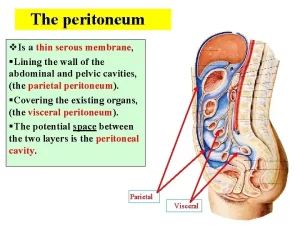
The peritoneum is a thin serous membrane that lines the walls of the abdominal cavity and invests the viscera. The parietal peritoneum lines the walls of the abdominal cavity. the anterior and posterior abdominal walls, the undersurface of the diaphragm, and the cavity of the pelvis.
The peritoneum
The visceral peritoneum is the continuation of the parietal peritoneum which leaves the posterior wall of the abdominal cavity to invest certain viscera. Between the parietal and visceral layers of the peritoneum is a potential space (the peritoneal cavity).
Abdominal viscera are either suspended in the peritoneal cavity by folds of the peritoneum (mesenteries) or are outside the peritoneal cavity, Organs suspended in the cavity are referred to as intraperitoneal; organs outside the peritoneal cavity, with only one surface or part of one surface covered by peritoneum, are retroperitoneal.
The peritoneal cavity is divided into two sacs
- Greater sac accounts for most of the space in the peritoneal cavity, beginning superiorly at the diaphragm and continuing inferiorly into the pelvic cavity, It is the part of the peritoneal cavity just behind the anterior abdominal wall.
- Lesser sac is a smaller subdivision of the peritoneal cavity posterior to the stomach and liver and is continuous with the greater sac through an opening, called the epiploic foramen of Winslow.
Peritoneal reflections in the sagittal section
To understand the extent of the greater sac, it is followed in a vertical direction. Following the peritoneum on the inner aspect of the anterior abdominal wall in an upward direction, a sickle-shaped fold of the peritoneum connects the anterior abdominal wall with the liver to the right of the median plane.
This ligament is called the falciform ligament. On the right side of the falciform ligament, the peritoneum continues on under the surface of the diaphragm. Then, it is reflected from under the surface of the diaphragm onto the superior surface of the liver. This reflection is called the upper layer of the coronary ligament.
The peritoneum passes from the upper surface of the liver to its anterior surface, then to its inferior surface. From the posterior part of the inferior surface, the peritoneum is reflected to the front of the right kidney and suprarenal gland. This reflection is called the lower layer of the coronary ligament. These two layers of coronary ligament bound the bare area of the liver on the posterior surface of the liver which has no peritoneal covering.
Following the upper and lower layers of the coronary ligament to the right shows that they meet at the apex of the bare area to form the right triangular ligament. From the front of the right kidney, the peritoneum passes to the front of the duodenum and right colic flexure. Just above the duodenum, the peritoneum passes medially in front of the inferior vena cava to form the posterior boundary of the epiploic foramen.
If the stomach is reflected away from the liver, a peritoneum fold (lesser omentum) is found connecting the liver at porta hepatis with the stomach. This is formed by two: layers. The two layers of the peritoneum separate to enclose the stomach. Then, they come close together at the greater curvature of the stomach to form a big peritoneal fold (greater omentum). The two layers of the greater omentum descend down and then reflected again upwards to be attached to the anterior border of the pancreas.
The peritoneum of greater sac passes down from the anterior border of the pancreas to form another peritoneal fold (transverse mesocolon) to enclose the transverse colon. Also, the peritoneum passes down to cover the inferior surface of the body of the pancreas, duodenum, and structures found in the posterior abdominal wall.
The peritoneum covering the posterior abdominal wall makes a reflection along the course of the superior mesenteric artery and encloses the small intestine (mesentery of the small intestine). The peritoneum continues down to continue into the pelvis to form the pelvic peritoneum.
Peritoneal reflections in the horizontal section
The peritoneum covers the inner aspect of the anterior abdominal wall and reflects in a horizontal pattern to form gastrosplenic, lienorenal, lesser omentum, and falciform ligaments of the liver.
Lesser sac
This is a diverticulum from the greater sac, it extends down behind the stomach as far as the transverse mesocolon and is bounded below the stomach by the greater omentum. The opening of the lesser sac (epiploic foramen) lies behind the free edge of the lesser omentum.
Boundaries of the epiploic foramen
- Posterior: The inferior vena cava lies immediately behind the posterior peritoneum.
- Superior: the inferior surface of the liver.
- Inferior: the first part of the duodenum.
- Anterior: free edge of the lesser omentum; in which the portal vein lies behind, the common bile duct in front, and the hepatic artery in front on the left of the bile duct.
Relations of the lesser sac
The lesser sac has two walls: anterior and posterior, and four borders: upper, lower, left, and right.
Anterior wall: It is formed by:
- The lesser omentum.
- The peritoneum covers the posterior surface of the stomach & first part of the duodenum.
- Anterior two layers of the greater omentum.
Posterior wall: Its lower part is formed by the posterior two layers of the greater omentum, while its upper part is formed by the peritoneum covering structures on the posterior abdominal wall which includes:
- Body of the pancreas.
- Diaphragm.
- Upper part of the abdominal aorta.
- Left kidney.
- Coeliac trunk and its branches.
- Left suprarenal gland.
Upper border: It is formed by reflection of the peritoneum between the liver and the diaphragm.
Lower border: It is formed by the inferior margin of the greater omentum where the anterior two layers become continuous with the posterior two layers.
Left border: formed by the gastrosplenic & lienorenal ligaments.
Right border: the epiploic foramen (foramen of Winslow).
You can download Science Online application on Google Play from this link: Science online Apps on Google Play
Constituents of the gastric juice, Gastric motility & types of movements occur in the stomach
Abdomen muscles, Blood Supply of Anterior Abdominal Wall & Rectus Sheath content
Temporal and infratemporal fossae contents, Muscles of mastication & Otic ganglion
Histological organization of pharynx, Structure & function of the esophagus
Mouth Cavity divisions, anatomy, function, muscles, Contents of Soft palate and Hard palate


