Ovarian hormones, Menopause, Functions of estrogen and progesterone in pregnancy
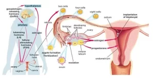
The ovaries produce eggs and hormones for menstruation and pregnancy, They are found on either side of the uterus, The major hormones secreted by the ovaries are oestrogen and progesterone, they are important in the menstrual cycle.
Ovarian hormones
Ovarian hormones are estrogens and progesterone.
Site of secretion
A. In nonpregnant females: Estrogen is secreted from the Graafian follicle and corpus luteum of the ovary, Progesterone is secreted during the latter half of each ovarian cycle by the corpus luteum. Minute amounts of female sex hormones are secreted by the adrenal cortex.
B. In pregnancy: Very large amounts of progesterone and estrogen are secreted by the placenta, Estrogens and progesterone are transported in the blood bound mainly with plasma albumin, The binding is so loose that they are rapidly released to the target tissue.
Function of estrogen
After puberty, the quantity of estrogens increases. At this time, the female sexual organs change from those of a child to those of an adult. Estrogen causes cellular growth and proliferation of tissues of sex organs and other tissues related to reproduction.
1. Effect on external sexual organs
The external genitalia enlarges, with deposition of fat in the mons pubis and labia majora and with enlargement of the labia minora.
2. Effect on genital organs
- Vagina: estrogens change the vaginal epithelium from a cuboidal into a stratified type which is more resistant to trauma and infection.
- Uterus: estrogens cause marked proliferation of the endometrial stroma and greatly increase the development of the endometrial glands. It increases the amount of uterine muscles and the blood flow to the uterus.
- Fallopian tube: estrogens cause the glandular tissues of the fallopian tubes to proliferate and increase the number and activity of ciliated epithelial cells that line the fallopian tubes.
3. Effect on the breasts
Development of the stromal tissues of the breasts and nipple. Growth of the duct system. Estrogens initiate the growth of the milk-producing apparatus. They increase the deposition of fats in the breasts giving it the external appearance of the mature female breast. It also causes pigmentation of the areola.
4. Effect on the skeleton
Estrogens cause increased osteoblastic activity. Therefore, at puberty, the growth rate becomes rapid for several years. They cause changes in body configurations. The woman has narrow shoulders and a broad hip, They cause early uniting of the epiphyses with the shafts of the long bones, After menopause, no estrogens are secreted, As a result, Osteoblastic activity decreases, Bone matrix decreases, and the deposition of bone calcium and phosphate decrease
5. Effect on protein metabolism
Estrogens cause a slight increase in total body protein (positive nitrogen balance). It has an anabolic effect due to its growth-promoting effect on the sexual organs, the bones and a few other tissues of the body.
6. Effect on metabolism and fat deposition
Estrogens increase the metabolic rate slightly. They also cause the deposition of fat in the subcutaneous tissues, buttocks, and thighs giving characteristics of the feminine figure.
7. Effect on the skin
Estrogens cause the skin to be soft and smooth and more vascular than normal.
8. Effect on electrolyte balance
Estrogen causes sodium and water retention by the kidney tubules.
Functions of progesterone
1. Effect on the uterus
Progesterone promotes secretory changes in the uterine endometrium during the latter half of the female sexual cycle, thus preparing the uterus for implantation of the fertilized ovum, Progesterone decreases the frequency of uterine contractions, thereby helping to prevent expulsion of the implanted ovum, and it makes cervical mucus thicker.
2. Effect on the fallopian tubes
It promotes secretory changes in the mucosal lining of the fallopian tubes. These secretions are for the nutrition of the fertilized ovum.
3. Effect on breasts
It promotes the development of the lobules and alveoli of the breasts, causing the alveolar cells to proliferate and enlarge to become secretory in nature.
4. Effect on electrolyte balance
Progesterone in very large quantities causes natriuresis.
5. Protein catabolic effect
Progesterone exerts a mild catabolic effect on the body’s protein. This effect is probably significant during pregnancy, when proteins must be mobilized for use by the fetus.
The menopause
At the age of 45-50 years, the sexual cycles usually become irregular and ovulation fails to occur during many of the cycles, The cycles cease and the female sex hormones diminish rapidly, The cause of menopause is “burning out of the ovaries FSH and LH are produced in large and continuous quantities because there is no ovarian hormone to inhibit them.
The loss of estrogens often causes marked physiological changes in the function of the body, including hot flashes characterized by extreme flushing of the skin, psychic sensations of dyspnoea, irritability, fatigue, anxiety, and occasionally various psychotic states.
Abnormalities of Secretion by the Ovaries
Hypogonadism
Before puberty: It results from poorly formed ovaries, lack of ovaries or genetically abnormal ovaries. In this condition, the manifestations are: The usual secondary sexual characteristics do not appear and the sexual organs remain infantile, Prolonged growth of the long bones: because the epiphyses do not unite with the shafts of these bones.
After puberty: When the ovaries of a fully developed woman are removed the following manifestations appear, The sexual organs regress to some extent so that the uterus becomes almost infantile in size, The vagina becomes smaller and the vaginal epithelium becomes thin and easily damaged, The breasts atrophy and become pendulous and the pubic hair becomes thinner, These same changes occur hi women after menopause.
Hypersecretion of the Ovaries
It is caused by tumors in the ovaries, These tumors secrete large quantities of estrogens, which exert the usual estrogenic effects, including hypertrophy of the uterine endometrium and irregular bleeding from this endometrium, Bleeding is often the first indication that such a tumor exists.
Hormones of pregnancy
In pregnancy, the placenta secretes the following hormones.
- Human chorionic gonadotropin.
- Estrogens.
- Progesterone.
- Human chorionic somatomammotropin.
- Relaxin.
Human Chorionic Gonadotropin
Coincidently with the development of the trophoblast cells from the early dividing ovum, the hormone is secreted into the fluids of the mother. The secretion of this hormone can first be measured 8 days after ovulation. Then the rate of secretion rises rapidly to reach a maximum of approximately 8 weeks after ovulation.
The functions of human chorionic gonadotropin are:
Human chorionic gonadotropin has very much the same molecular structure and function as the luteinizing hormone.
- Prevention of the normal involution of the corpus luteum.
- It causes the corpus luteum to secrete larger quantities of progesterone and estrogen which cause the endometrium to continue growing and to store large amounts of nutrients.
- It exerts interstitial cell-stimulating effects on the testis, thus resulting in the production of testosterone in the male fetus.
Functions of estrogen in pregnancy
- It stimulates the continuous growth of the myometrium.
- It increases the growth of the ductal system of the breast.
- It stimulates the enlargement of the external genitalia.
- It relaxes the various pelvic ligaments.
Functions of progesterone in pregnancy
- Progesterone causes decidual cells to develop in the uterine endometrium for the nutrition of the early embryo.
- Progesterone decreases the contractility of the gravid uterus, thus preventing uterine contractions from causing spontaneous abortion.
- it helps in the development of the zygote prior to implantation.
- It increases the secretions of the fallopian tubes and uterus to provide appropriate nutritive matter for the developing zygote
- Progesterone prepares the breasts for lactation.
Human chorionic somatomammotropin
A new placental hormone which is a protein secreted about the fifth week of pregnancy and increases progressively. Its functions are:
- It causes partial development of the breasts.
- It has a lactogenic activity like prolactin.
- It has weak actions similar to those of growth hormone, Causing deposition of protein in the tissues.
- It causes sodium, potassium, and calcium retention.
- It causes decreased utilization of glucose by the mother, thereby making larger quantities of glucose available to the fetus.
- It promotes the release of free fatty acids from the fat stores of the mother, thus providing an alternative source of energy for her metabolism.
Relaxin Hormone
It is secreted by the corpus luteum of pregnancy and by decidual cells. The hormone. relaxes pelvic bones and ligaments, inhibits myometrial contractions and softens the cervix.
Other Maternal Hormonal Changes
1. Anterior pituitary gland: Maternal growth hormone secretion in response to various stimuli decreases during pregnancy, probably because its anabolic functions are carried out by human chorionic somatomammotropin and/or placental growth hormone variant. Maternal LH and FSH secretion also is suppressed by the high levels of estrogen, progesterone, and inhibin from the corpus luteum and later the placenta.
2. Adrenal cortex: Glucocorticoid secretion is increased throughout pregnancy. The enhanced glucocorticoid activity may contribute to maternal adipose tissue gain and to mammary gland development. Aldosterone secretion increases significantly throughout pregnancy, this contributes to the positive sodium balance that is needed to maintain a high total maternal plasma volume and to build the extracellular fluid of the fetus.
3. Thyroid glands: total plasma thyroid hormones are elevated. The thyroid hormones stimulate the metabolic activities in the mother.
4. Insulin secretion, in response to meals increases after the third month of secretion This secretion reaches its peak during the last trimester coinciding with the peak of plasma human somatomammotropin. Because maternal sensitivity to insulin is greatly diminished during this period, insulin hypersecretion may be compensatory.
5. Basal glucagon levels do not change significantly.
6. Calcium absorption from the diet increases during pregnancy and offsets the continuing drain created by the growing fetal skeleton. This increase in calcium absorption is accounted for by increased maternal levels of the active metabolite of Vitamin D.
You can download Science online application on google play from this link: Science online Apps on Google play
Female reproductive system organs, functions, anatomy & Histological structure of the uterus
Ovary function, location, anatomy, structure, Oogenesis, and maturation of primary oocyte
Fallopian tubes function, location, anatomy & Histological structure of vagina
Female mammary glands (breast), signs of breast cancer & Mammary gland abnormalities


