Neuro-Muscular Junction properties, Functions & types of skeletal muscles
Neuro-Muscular Junction (Motor End-plate) is a specialized nerve ending (effector, motor) by which a motor nerve fiber ends in a skeletal muscle. Skeletal muscles are innervated by large myelinated nerve fibers, originating from the motor neurons of the cranial nerves and anterior horn cells of the spinal cord.
Histological structure of motor endplate
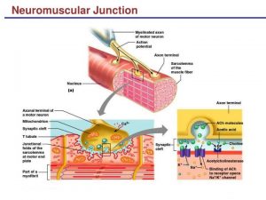
The site of the junction between the nerve fiber and the skeletal muscle fiber is formed of:
Axon terminal: near the sarcolemma, the myelinated motor nerve fiber loses its myelin sheath and divides into several branches forming axon terminals, which terminate in contact with the sarcolemma by the epilemmal ending. Thus, there is no cytoplasmic continuity between the nerve terminal and muscle fiber. The axon terminal contains a large number of mitochondria and synaptic vesicles containing the neurotransmitter, acetylcholine.
Synaptic cleft is the space between the axon terminal and the sarcolemma of the skeletal muscle fiber. In this cleft, a high concentration of acetylcholine esterase enzyme is found.
Postsynaptic sarcolemma: under the axon terminal, the sarcolemma is corrugated forming junctional folds to increase the surface area of sarcolemma exposed to the neurotransmitter, The postsynaptic sarcolemma contains numerous receptors for acetylcholine.
Sole plasm is the sarcolemma under the postsynaptic sarcolemma. It is characterized by being slightly elevated with many nuclei. It appears granular as it contains numerous mitochondria, abundant glycogen granules, and ribosomes. Myofibrils are absent in the sole plasm.
Physiology of skeletal muscle
Muscles are machines for converting stored chemical energy into mechanical energy (work) and heat. Muscles constitute 50% of the total body weight; contraction is the main function of the muscles. The individual muscle fibers are the ” building blocks” of the muscular system, skeletal muscles end in tendons, and the muscle fibers are paralleled so that the force of contraction of the units is additive. There are approximately 640 skeletal muscles in the human body.
Functions of skeletal muscles
- They move the body as a whole or part of it e.g. one limb.
- They maintain the body posture by their tonic contraction and muscle tone.
- Generating heat by their contraction, which is vitally important in maintaining normal body temperature.
- Stabilize and strengthen the joints of the skeleton by their contraction.
Types of skeletal muscle fibers
Skeletal muscles contain mainly two types of muscle fibers.
a. Slow (red) muscle fibers (type I)
This type of muscle fibers is characterized by:
- Thin fibers contain large amounts of myoglobin, which combines with oxygen and stores it until needed.
- Numerous blood capillaries supply extra amounts of oxygen (red fibers).
- A greatly increased number of mitochondria to support high levels of aerobic oxidation.
- These muscles have a relatively longer latent period, contract slowly, and fatigued slowly.
- These muscles are adapted for long slow contractions which maintain the body posture against gravity e.g. back muscles and soleus muscle and also, are great at helping athletes run marathons and bicycle for hours.
b. Fast (pale) muscle fibers (type II)
This type of muscle fibers is characterized by:
- Thick fibers for great strength of contraction.
- Extensive sarcoplasmic reticulum for rapid release of calcium ions to initiate contraction.
- Large amounts of glycolytic enzymes for rapid release of energy by the glycolytic process (anaerobic oxidation).
- Myoglobin is absent; there are few blood capillaries (pale fibers) and few mitochondria.
- These muscles have a short latent period, contract rapidly, and fatigued easily.
- They are adapted for fine skilled movements e.g. extraocular muscles and some hand muscles.
- Because fast twitch fibers use anaerobic metabolism to create fuel, they are much better at generating shorts bursts of speed that can help a sprinter since he needs to quickly generate a lot of force.
In the human body, individual skeletal muscles tend to have a mixture of the two twitch types. The third type of muscle fibers (intermediate type; fast oxidative) are present. These fibers share the characteristics with each of the other two types i.e. they have high ATPase activity like the fast (anaerobic) fibers and high oxidative capacity like the slow (aerobic fibers).
Nerve supply of skeletal muscle
Skeletal muscles are nerve-operated and under the control of the CNS, they contract only when their motor nerve is intact, connecting them to the CNS. Motor nerve fibers reach the skeletal muscle from the anterior horn cells in the grey matter of the spinal cord. Degeneration of muscle with complete loss of function or paralysis occurs if its nerve supply is cut.
Motor unit (The all or none rule)
Each anterior horn cell together with its axon and the number of muscle fibers it supplies (about 150-30 muscle fibers) is called a motor unit. When the anterior horn cell is stimulated, all its muscle fibers contract (each motor unit obeys the all or non-rule), not all the motor units in a given muscle, contract at the same time. The number of active motor units increases, when the intensity of stimulation of the muscle increases.
Changes occurring in a muscle as a result of activity
When skeletal muscle is stimulated either directly or indirectly through its motor nerve supply, the following changes occur:
- Electrical changes.
- Excitability changes.
- Mechanical changes.
- Chemical or metabolic changes.
- Heat production or thermal changes.
Electrical changes in a skeletal muscle
They are the same as in the nerve with some differences:
- Resting membrane potential in the skeletal muscle fibers is -90 mv (in small nerve fibers -70 mv).
- The magnitude of the action potential is 130 millivolts (-90 to +40 mv), in the thick myelinated nerve fibers, it is 105 mv (-70 to +35).
- The duration of the action potential in skeletal muscle is 1-5 msec (in the thick myelinated nerve fibers, it is (2-4 msec).
- The velocity of conduction of excitation wave is from 3-5 meters/second, (in the thick myelinated nerve fibers, it reaches up to 120 meters/ sec).
- The muscle spike potential precedes the muscle contraction by about 2 msec.
Neuro-muscular junction (motor end plate)
Mechanism of neuro-muscular transmission
- Upon the arrival of an action potential at the axon terminal, voltage-dependent calcium channels open, and Ca++ ions flow from the extracellular fluid into the membrane of the nerve terminals.
- The influx causes neurotransmitters- containing vesicles to attach Ca++ to the membrane of the nerve fiber.
- Fusion of the vesicular membrane with the nerve cell membrane results in the emptying of the vesicle’s contents; acetylcholine, into the synaptic cleft, a process known as exocytosis.
- Acetylcholine diffuses into the synaptic cleft and binds to the acetylcholine receptors at the motor end-plate.
- These receptors are ligand-gated ion channels, and when they bind acetylcholine they open, allowing a rapid influx of Na+ ions to the interior of the muscle fiber, to excite the generation of an action potential.
End plate potential (EPP)
Sudden entry of sodium ions into the muscle fiber decreases the membrane potential in the local area of the end plate, creating a local potential called the end plate potential (partial depolarization of the membrane).
The end plate potential is a local unpropagated potential when it reaches a certain value called threshold potential it fires the potential on both sides of the motor end plate, along the sarcolemmal membrane, leading to muscular contraction.
Fate of acetylcholine (ACh)
Acetylcholine is rapidly destroyed (after one millisecond) from its release, by the acetyl cholinesterase enzyme in the cleft itself. This short time is sufficient for acetylcholine to excite the muscle fibers. The rapid hydrolysis of acetylcholine prevents re-excitation of the muscle fiber after recovery from the previous action potential.
Properties of neuromuscular transmission
- Unidirectional: Neuromuscular transmission occurs in one direction from the nerve to the muscle and not in the opposite direction.
- Delay: There is some delay of about 0.5 msec. in neuromuscular transmission. This time is used for release of acetylcholine and its binding to the receptors at the outer surface of the muscle membrane. Depolarization occurs and EPP is created till it reaches the firing level and an action potential is generated at the muscle fiber membrane.
- Fatigue: The neuromuscular junction is the first site in the neuromuscular system which suffers from fatigue.
Myasthenia gravis
- Myasthenia gravis is a long-term neuromuscular autoimmune disease that results from antibodies that block or destroy nicotinic acetylcholine receptors at the junction between the nerve and muscle.
- This prevents nerve impulses from triggering muscle contraction
- It leads to varying degrees of skeletal muscle weakness. The most commonly affected muscles are those of the eyes, face, and swallowing. It can result in double vision (diplopia), drooping eyelids, trouble talking, and trouble walking.
- It is characterized by tiredness and progressive paralysis of muscles; death commonly occurs due to failure of respiratory muscles.
- Myasthenia gravis is generally treated with medications known as acetylcholinesterase inhibitors such as neostigmine and pyridostigmine.
- Drugs such as neostigmine., inhibit the action of acetylcholine esterase and allow the accumulation of adequate amounts of acetyl choline for a long time to stimulate the remaining receptors.
Muscles types, Skeletal muscle parts, Lymphatic system structure & function
Muscular system, Structure of skeletal muscle, Muscles properties & functions
Muscular tissue types, function, structure, definition & anatomy
Excitability changes in skeletal muscle fibers during activity & Causes of muscle fatigue


