Masses in Right iliac fossa, lumbar region, and umbilical region
A right iliac fossa (RIF) mass can arise from various structures in that region, including the gastrointestinal tract, genitourinary system, and lymphatic tissue. The differential diagnosis depends on the patient’s age, sex, and associated symptoms.
Right iliac fossa
1- Appendicular Mass
- It is a smooth, firm, tender mass in the right iliac fossa.
- It is not mobile. It does not move with respiration.
- It is resonant on percussion. It is a well-localized mass with distinct borders.
2- Appendicular Abscess
It is a smooth, soft, tender, and dull mass in the right iliac fossa with indistinct borders.
3. Carcinoma of the caecum
- It is a nodular, hard, mass in the right iliac fossa.
- It does not move with respiration.
- It is mobile but mobility may be restricted once it gets adherent to the psoas muscle.
- Mass is resonant or there is impaired resonance on per cussion.
- Often features of intestinal obstruction may be present.
4- Ileocaecal Tuberculosis
- Mass in the right iliac fossa which is smooth, hard, resonant, and nontender.
- It does not move with respiration and has restricted mobility.
- The caecum may be pulled up to lumbar region due to fibrosis.
5- Amoeboma
- History of dysentery with pain in the right iliac fossa.
- Smooth, hard, well-defined mass in the right iliac fossa which is nonmobile.
- It may or may not be tender.
6- Psoas Abscess
- It is a localized, smooth, soft, nonmobile mass in the right iliac fossa.
- Psoas spasm (flexion of the hip joint) is typical.
- The spine may show gibbus, tenderness, and paraspinal spasm. Spinal movements will be restricted.
Mass in the lumbar region
Palpable Kidney Mass
- There is fullness in the loin which is better observed in a sitting position.
- Mass moves with respiration. It is vertically placed.
- It is bimanually palpable. It is ballotable.
- The renal angle is dull on percussion (normally it is resonant due to the colon).
- There is a band of resonance in front due to the reflected colon.
- It does not cross the midline.
Conditions Where Kidney Gets Enlarged
Hydronephrosis: It is smooth, soft, lobulated, nontender mass, and nonmobile.
Pyonephrosis:
- History of throbbing pain in the loin, pyuria, and fever with chills.
- It is smooth, soft and tender kidney mass, nonmobile.
Polycystic kidney:
- History of loin pain and haematuria.
- Hypertension, anaemia and features of renal failure.
- Usually bilateral. But one side can present early than on the other side.
- Lobulated smooth surface.
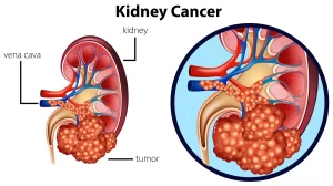
Renal cell Carcinoma):
- History of mass in the loin, haematuria, fever, and dull pain.
- Mass is nodular and hard.
- It does not cross the midline.
- Initially mobile; eventually it infiltrates gets fixed and becomes nonmobile.
Mass from the Ascending Colon on the Right Side or Descending Colon on the Left Side
- History of altered bowel habits with decreased appetite and weight.
- Mass is nodular, hard which does not move with respiration, and is not ballotable
- It is resonant or there is impaired resonance on percussion.
- The renal angle is resonant.
- Proximal dilated bowel may be palpable.
Adrenal Mass
- It is nodular and hard.
- It does not move with respiration.
- It is not mobile and often crosses the midline.
- It is felt on deep palpation.
- It is resonant in front.
- It is not ballotable.
Retroperitoneal Tumours
- They are not mobile, or resonant, and do not fall forward in knee-elbow position.
- They are deeply placed masses which are usually smooth and hard.
- They may be retroperitoneal sarcomas or teratomas or lymph node mass.
Retroperitoneal Cysts
They are smooth and soft with the same features as retroperitoneal tumours.
Cystic lesions in the abdomen
- Mucocele/empyema of gallbladder
- Pseudocyst of the pancreas.
- Hydatid cyst of liver
- Congenital nonparasitic cyst of the liver.
- Hydronephrosis.
- Mesenteric cyst.
- Ovarian cyst.
- Omental cyst.
- Aneurysm.
- Retroperitoneal cyst.
- Cyst adenocarcinoma of ovary.
- Loculated ascites.
Mass in the umbilical region
Lymph node mass
secondaries (primary from GIT, testis, ovary, melanoma)/lymphoma/tuberculosis
Mesenteric Cyst
Tillaux triad:
- Soft intra-abdominal umbilical mass.
- Mobile in the direction perpendicular to the attachment of the mesentery.
- Resonant mass.
May precipitate intestinal obstruction, volvulus.
Omental Cyst
- It is smooth, soft, and nontender.
- It moves with respiration. It is mobile in all directions.
- It is dull on percussion.
Small Bowel Swellings
- Small bowel lymphomas.
- Small bowel carcinomas.
- Intussusception.
Intussusception
- Mass in the umbilical region usually towards the left and above the umbilicus,
- Occasionally towards the right side.
- Mass is intra-abdominal which is sausage-shaped, with concavity towards the umbilicus, well-defined, smooth, firm, and mobile.
- Mass does not move with respiration.
- Mass contracts under palpating fingers.
- Often mass disappears and reappears.
- Mass is resonant or there is impaired resonance on percussion.
- Red currant gellyl stool with features of intestinal obstruction may be present.
Investigations for Mass Abdomen
- Hematocrit, liver function tests, renal function tests, stool/ urine examination.
- Ultrasound abdomen.
- Endoscopies-gastroscopy-colonoscopy-ERCP-MRCP.
- Barium studies-Barium meal-Barium enema-Barium meal – Follow through.
- CT scan – MRI.
- Endosonography.
- Ascetic tap.
- Diagnostic laparoscopy.
- U/S guided/CT guided biopsy.
- IVU/RGP/Cystoscopy/Isotope renogram.
- Exploratory laparotomy.
A hard mass in the abdomen is commonly malignant. Firm mass may be tuberculous, lymphoma, or many benign conditions. Soft masses are hydronephrosis, pseudocyst, mesenteric cyst, omental cyst, and loculated ascites. In tuberculosis abdomen may be doughy due to thickened parietal peritoneum or omentum.
It is difficult to elicit fluctuation in the mass abdomen as the mass cannot be fixed properly. Plane of the swelling should be checked by leg raising/head raising test/Valsalva manoeuvre/knee elbow test. Bimanual palpation, ballotability, renal angle inspection, palpation, and percussion should be done in case of renal mass
Intrinsic mobility should be checked. Different mobilities/movements are gallbladder shows side to side; stomach lateral; ovarian mass-all over, mesenteric cyst-right angle to line of mesentery; transverse colon mass-vertical: small bowel mass all over, appendicular mass/pancreatic mass/ nodal mass (Para-aortic)/retroperitoneal mass do not show any mobility; cyst adenocarcinoma of pancreas may show false mobility (tree top mobility).
Percussion is very important method of examination to find out the anatomical plane of the mass. Mass in front of the bowel like liver/spleen/gallbladder/parietal mass is dull on percussion. Mass from the bowel is resonant on percussion like from the stomach, small bowel and colon. Retroperitoneal masses like cysts, sarcoma, nodal masses, aneurysm, pancreatic mass are resonant on percussion.
Succussion splash and auscultopercussion tests are done for gastric outlet obstruction in pyloric stenosis. Digital examination of the rectum (P/R) and left supraclavicular examination for nodes is a must. PR is done to see Blumer shelf secondaries in the rectovesical pouch. Pervaginal examination should be done in pelvic mass in females.
The bladder should be emptied while examining the pelvic mass. The bimanual examination is done often under general anesthesia in case of a pelvic mass. Auscultation for bruit depends on the condition and location of mass over the epigastrium, over the liver.
Masses that may appear and disappear
- Pseudocyst of pancreas (communicating).
- Hydronephrosis (intermittent).
- Choledochal cyst.
- Intussusception.
You can subscribe to Science Online on YouTube from this link: Science Online
Gastroesophageal Reflux Disease, Complications of GERD and Barrett’s oesophagus
Esophagus diseases, Dysphagia causes, Achalasia, and Symptomatic Diffuse Esophageal spasm
Pharynx function, anatomy, location, muscles, structure, and Esophagus parts
Tongue function, anatomy, and structure, Types of lingual papillae, and Types of cells in taste bud
Mouth Cavity divisions, anatomy, function, muscles, Contents of Soft palate and Hard palate
Temporal and infratemporal fossae contents, Muscles of mastication and Otic ganglion


