Intestine development, congenital anomalies, anatomy, structure and function
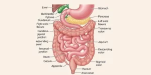
The intestine extends from the lower end of your stomach to your anus, the lower opening of the digestive tract. It is called the bowel or bowels, Food and the products of digestion pass through the intestine, which is divided into two sections called the small intestine and the large intestine, Most absorption of nutrients and water happens in the intestine. The intestine absorbs nutrients and vitamins, It is a long, continuous muscular tube, and it is part of the gastrointestinal (GI) tract.
Development of Intestine
Except Duodenum and Hindgut
Origin: Endoderm of midgut → mucosa & glands.
Splanchnic secondary mesoderm → submucosa & musculosa and serosa.
Stages of development
1- Preherniation stage:
- Straight midgut → midgut loop → cranial limb and caudal limb.
- Cranial limb → small intestine (jejunum + proximal part of ileum).
- Caudal limb → large intestine (distal part of ileum + caecum +appendix + ascending colon + right 2/3 transverse colon).
2- Herniation stage:
Causes:
- Small abdominal cavity.
- Large liver and kidney.
- Extraembryonic coelom in the primitive umbilical cord.
Site: Extraembryonic coelom of the umbilical cord.
Rotation 90° anticlockwise → cranial limb → right limb (small intestine) & caudal limb → left limb (large intestine).
3- Posthemiation stage:
- Reduction of small intestine first, large intestine second, and caecum last.
- Rotation 180° Anticlockwise → right limb → left (small intestine) and left limb → right (large intestine).
- Proliferation followed by recanalization of the endoderm of the intestine occurs.
Congenital anomalies
- Congenital umbilical hernia: It is due to the presence of a defect in the anterior abdominal wall → herniation of loop of the intestine.
- Exomphalos: It is due to failure of a loop of the intestine to return back to the abdominal cavity → loop of the intestine coming from the base umbilicus covered by amnion.
- Meckel’s diverticulum: It is due to the persistence of the small pouch of the yolk stalk. Length: 2 inches * Distance from caecum: 2 feet. Incidence: 2% of people.
- Congenital umbilical (fecal) fistula due to the persistence of all yolk stalk.
- Rotation clockwise → transposition.
- Intestinal atresia: due to failure of recanalization of the intestine.
- Intestinal stenosis: due incomplete recanalization.
- Duplication of the intestine: duplication of segment of the intestine due to abnormal recanalization.
Development of duodenum
Origin:
- Endoderm of foregut → mucosa of 1st + ½ second part.
- The endoderm of midgut → mucosa of ½ second +3rd+ 4th parts.
- Splanchnic 2ry mesoderm → submucosa & musculosa.
Development:
- It starts as a vertical tube formed of the caudal part of the foregut and the cranial part of the midgut.
- Unequal growth of the walls & traction of Treitz ligament (from the duodenojejunal junction to the left crus of the diaphragm) leads to:
- C-shaped loop projecting anteriorly.
- The ventral opening of the bile duct will be shifted posteromedial.
Rotation of the duodenum to the right (clockwise) leads to:
- The ventral convex margin → right.
- The right surface→ posterior.
The proliferation of the endoderm occurs followed by recanalization.
Congenital anomalies
- Duodenal atresia: due to non canalization. It is usually associated with poly hydroamnios.
- Duodenal stenosis: due to partial canalization.
- Congenital intestinal obstruction: due to traction of Treitz ligament on the duodenojejunal junction.
- Duplication of the duodenum: due to abnormal recanalization.
Development of rectum and upper ½ of the anal canal
Origin:
- Endoderm of hindgut → mucosa & its glands.
- Splanchnic secondary mesoderm → submucosa & musculosa.
Development:
- The cloaca (dilated caudal part of hindgut) is divided by a cloacal septum (wedge-shaped mesoderm between allantois and hindgut) which grows towards the cloacal membrane into:
- A dorsal part (anorectal canal).
- A ventral part (primitive urogenital sinus).
- The cloacal membrane is also divided into anal and urogenital membranes.
- The anorectal canal gives the rectum and upper 1/2 of the anal canal.
- The rectum will be convoluted due to unequal growth of its walls.
Development of lower half of anal canal
Origin:
- Ectoderm → Stratified squamous epithelium.
- Splanchnic secondary mesoderm → submucosa, musculosa, and anal sphincters.
So, its upper is endodermal and its lower half is ectodermal.
Development:
- Mesenchyme around the anal membrane proliferate producing elevations of the surface ectoderm called anal hillocks.
- The anal membrane is now located at the bottom of an ectodermal depression called proctodium (primitive anal canal) → lower of anal canal.
- The anal membrane ruptures leaving remnants as anal valves and the two halves of the anal canal are continuous with each other.
Congenital anomalies
- Fistulae formation: (Rectovaginal, rectouretheral, rectovesical, anouretheral in males and anovaginal fistula in females) due to incomplete development of cloacal septum.
- Rectal atresia: due to either abnormal canalization or defect in blood supply causing focal atresia.
- Imperforate anus: due to failure of rupture of anal membrane. A layer of tissue separates the anal canal from the exterior.
- Anal stenosis: due to dorsal deviation of the cloacal septum as it grows caudally → small anal canal.
- Anal agenesis: it terminates blindly. It associates with a fistula between the rectum and urinary bladder, urethra in males, or vagina in females.
You can download Science online application on Google Play from this link: Science online Apps on Google Play
Intestinal motility, Intestinal movements types & Factors affecting Rate of intestinal absorption
Small intestine constituents, Regulation, function and Digestion in the small intestine
Small intestine function, anatomy, parts, Arterial supply of the duodenum, midgut & hindgut
Large Intestine function, parts, length, anatomy & Relations of the rectum


