Histological organization of vestibular apparatus, Hearing importance and body Equilibrium
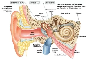
The inner ear is one of the special sense organs concerned mainly with hearing and maintaining the body equilibrium. The Inner ear is a fluid-filled cavity that consists of osseous and membranous labyrinths:
Osseous (Bony) labyrinth
It consists of a series of connected irregular spaces within the petrous portion of the temporal bone, It is filled with the perilymph, It is formed of the following structures:
- Cochlea, which lies anteriorly.
- Vestibule, which lies centrally. The vestibule is the most expanded part of the bony labyrinth, from it arise both the cochlea and the semicircular canals.
- Three bony semicircular canals, which lie posteriorly.
Membranous labyrinth
It is formed of a continuous series of sacs and channels that are suspended inside the bony labyrinth. The parts of the membranous labyrinth are filled with the endolymph and are surrounded by the perilymph lying between the membranous and bony labyrinths.
The membranous labyrinth is lined by simple epithelium which, in certain sites, becomes thickened by the neuroepithelial cells that are specialized for either auditory or vestibular function. The membranous labyrinth is formed of:
- Cochlear duct fitting in the bony cochlea and containing the organ of Corti.
- Utricle and saccule fitting in the bony vestibule and containing the two maculae one in the utricle and the other in the saccule.
- Three membranous semicircular ducts fitting in the three bony semicircular canals and containing the three cristae ampullaris.
Vestibular apparatus
A. The utricle and sacculer
They are two membranous sacs lying inside the bony vestibule and are joined by γ-shaped endolymphatic duct. The maculae (sacculi & utriculi) are present in the saccule and utricle of the vestibule.
Macula sacculi and macula utriculi (The otolith organ)
These are neuroepithelial thickenings in the membranes of the utricle and saccule. They are responsible for the sensation of linear movement of the head. The maculae (sacculi & utriculi) are similar in their structure and are composed of:
I. Hair cells: these are specialized neuroepithelial cells of 2 types:
Type I hair cells
- They are flask-shaped cells and their free surface has numerous rows of stereocilia and a single long, non-motile kinocilium, located at one side of the cell.
- The stereocilia are arranged in rows of decreasing length, with the longest row lie adjacent to the kinocilium.
- Afferent fibers of the vestibular division of the VIll nerve arising from the vestibular ganglion cells, penetrate between the supporting cells and each forms a cup-shaped nerve ending, entirely surrounding the rounded base of the hair cell.
Type Il hair cells
- They are columnar in shape and have similar apical stereocilia and a single kinocilium.
- They differ in the form of their innervations. The afferent nerves form several small bouton nerve endings around the basal part of the cell.
II. Supporting cells: they are columnar cells disposed between the hair cells. They have few microvilli on their free surface.
III. The otolithic membrane:
- It is a covering of a thick gelatinous layer of proteoglycans with surface deposits of calcium carbonate crystals; otoliths (otoconia).
- Stereocilia and kinocilia of hair cells are embedded into this membrane.
B. The semicircular canals and ducts
There are three bony semicircular canals (SSCs), the lateral (horizontal), the anterior superior, and the posterior. They are arranged at planes that are perpendicular to each other. Each canal has a dilatation at its base called the ampulla. The three membranous ducts have the same general form as the bony canals assuming the same course and having the same dilatations (ampullae). Both ends of each semicircular duct open in the utricle of the vestibule.
Crista ampullaris
The semicircular membranous ducts at the level of their ampullae show neuroepithelial thickenings called the cristae ampullares. Their structure is very similar to that of maculae. They are responsible for the sensation of rotation (angular acceleration) of the head. The crista ampullaris is composed of:
- Hair cells: two types are present, type I and type Il hair cells, similar to those of maculae. They are also innervated by the vestibular division of the VIII nerve.
- Supporting cells.
- The cupula: It is a gelatinous layer of proteoglycans where the stereocilia and kinocilia of the hair cells are embedded. It is conical in shape and is not covered by otoliths.
You can subscribe to Science Online on YouTube from this link: Science Online
Ear anatomy, structure, function, Relations of middle ear & Action of Auditory muscles
Physiology of equilibrium, Hearing, ear balance, Function & Stimulants of Semicircular canals
Auditory system structure & function, Auditory apparatus, cochlea & cochlear duct
Hearing, Transmission of sound waves in Cochlea, Functions of Cochlea & Auditory pathway


