Eyes structure, Histological organization of the fibrous and vascular coast of the eye
Eyes are important in seeing, Sight allows us to perceive movement, and vision allows us to make assessments about that movement, They keep us safe, Sight and vision offer awareness of the dangers around us, sight is the most important sense for safety & self-preservation, Protecting your eyes and your sight is important to avoid harm.
Eye structure
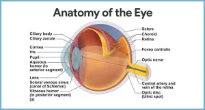
The eye is a highly-developed photosensitive organ that provides the sense of sight. It is composed of:
- The eyeball is located within the bony orbit.
- The accessory structures of the eye: the conjunctiva, the eyelids, and the lacrimal apparatus.
The eyeball is formed of 3 coats:
- Outer fibrous coat (Corneo-scleral coat): it is formed of the cornea and the sclera.
- Middle vascular coat (Uveal tract): it is formed of the choroid, the ciliary body, and the iris.
- Inner nervous coat: the retina.
The fibrous coat of the eye
A. The cornea
It is the transparent avascular anterior one-sixth of the external coat. Histologically, it consists of five layers:
1. Corneal epithelium: It is a non-keratinized stratified squamous epithelium that is characterized by the following:
- It is formed of 5-6 layers of epithelial cells resting on a straight basement membrane. The basal layer shows mitotic figures, denoting a high regenerative capacity of the cornea.
- The surface corneal cells show microvilli that protrude into the pre-corneal tear film (a protective layer of lipid and glycoprotein).
- Rich in free nerve endings, thus extremely sensitive to touch and pain.
2. Bowman’s membrane: it is the anterior layer of the corneal stroma. It acts as a protective barrier against trauma and bacterial invasion. However, it does not have a regenerative capacity. Therefore, if this layer is destroyed, healing always leaves a corneal scar and opacity.
3. Corneal stroma (substantia propria):
- It forms 90% of the thickness of the cornea.
- It is formed of successive lamellae of collagen fibers.
- Between these lamellae, flattened fibroblasts are present called corneal corpuscles.
4. Descemet’s membrane: it is the thick basement membrane of the corneal endothelium.
5. The corneal endothelium: the cornea is lined internally by simple squamous epithelium. Cells of this layer are involved in the metabolic exchange between aqueous humor and cornea.
The histological features that contribute to corneal transparency
The corneal surface epithelium with its relatively few layers and the straight basement membrane. The avascular corneal stroma i.e. no capillaries, no lymphatics. The cornea is nourished from the aqueous of the anterior chamber and by diffusion from blood vessels of the sclera. The uniform spacing of the parallel collagen fibers within each lamella of the corneal stroma. The collagen fibers in the alternating lamellae are oriented at right angles to one another.
B. The sclera
It is the opaque posterior five-sixths of the external coat forming the white of the eye. It consists of dense, irregular connective tissue (type I collagen fibers and elastic fibers. Corneo-scleral junction (The eye limbus): This is the transition zone between the transparent cornea and the white opaque sclera.
At the limbus, the following changes occur:
- The epithelium of the cornea is continuous with that of the bulbar conjunctiva.
- Bowman’s membrane ends abruptly and is replaced by the subconjunctival connective tissue.
- The regular lamellae of the corneal stroma merge with the irregular collagen bundles of the sclera.
- Descemet’s membrane and corneal endothelium change into the discontinuous layer of fibroblasts and melanocytes on the anterior surface of the iris.
The vascular coat of the eye
A. The choroid
This is a highly vascular and pigmented layer adjacent to the inner surface of the sclera. This layer is responsible for supporting the retina, absorbing excessive light, and providing the retina with blood supply and nutrients.
B. The ciliary body
This is the anterior expansion of the choroid forming a ring-like thickening that encircles the eye at the limbus. In the transverse section, it forms a triangle with its small base facing the anterior chamber and its outer surface faces the sclera. The inner surface, towards the posterior chamber, shows the ciliary processes that give rise to zonule fibers. These fibers are attached to the lens capsule keeping it in position.
Histologically, the ciliary body is made up of:
1. Ciliary epithelium: it consists of a double layer of low columnar epithelial cells that are covering the ciliary processes and are arranged as:
- Non-pigmented inner layer of cells i.e. towards the posterior chamber.
- The pigmented outer layer of cells l.e. towards the ciliary stroma.
The ciliary epithelium has the following functions:
- Secretion of aqueous humor
- Secretion of zonule fibers.
- Formation of the blood-aqueous barrier: through the tight junctions between the apices of the non-pigmented ciliary epithelial cells.
2. Ciliary stroma: it consists of loose connective tissue rich in blood vessels and melanocytes.
3. Ciliary muscles: they form the bulk of the ciliary body. There are three functional groups of smooth muscle fibers, their contraction controls the:
- Drainage of aqueous humor.
- Accommodation for near & far vision.
C. The iris
It is responsible for the color of the eye. It lies in front of the eye lens, covering its periphery and leaving its central part uncovered to allow the passage of light through an opening (pupil). The iris is formed of pigmented, highly vascular connective tissue containing the smooth muscles of the iris (sphincter & dilator pupillae).
Circulation of the aqueous humor
- The aqueous humor is produced by the ciliary epithelium, passes from the posterior chamber of the eye into the interior chamber through the opening of the pupil between the iris and lens to reach the limbus.
- At the angle of the limbus, aqueous passes through the openings of the trabecular meshwork of the limbus and then exits through the canal of Schlemm (scleral venous sinus).
- The canal of Schlemm is an irregular channel lined with endothelium and is running circularly around the limbus. It collects the aqueous fluid from the anterior chamber for its drainage Into the episcleral veins.
Excessive secretion or interference with the drainage of aqueous fluid causes an increase in intraocular pressure (glaucoma).
The eye lens
This is a transparent biconvex flexible disc situated immediately behind the pupil. It is formed of tightly-packed elongated lens fibers. The eye lens is enveloped by the lens capsule into which the zonule fibers of the ciliary processes are inserted, forming the suspensory ligaments of the lens. According to the tension of the zonule on the lens substance, controlled by ciliary muscles, lens curvature would change to serve as accommodation for near and far vision.
Vitreous body
It is a transparent refractile gel that fills the cavity of the eye behind the lens.
You can subscribe to Science Online on YouTube from this link: Science Online
You can download the Science Online application on Google Play from this link: Science Online Apps on Google Play
Visual system, Bony orbit anatomy, contents & Nerves, Muscles & movements of Eyeball
Retina function, anatomy, layers and accessory structures of the eye
Sense of vision, Refractive media of the eye, Corneal reflex & Pupillary pathways
Retinal receptors, Photochemistry of vision, Steps of phototransduction in rods & Retinal output
Nystagmus types, causes, symptoms Development of CNS & Abnormalities of Spinal cord


