Chimerism Cause, Types of structural chromosomal abnormalities and Indications for Chromosomal Analysis
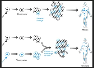
A chimer results from the fusion of 2 different (twin) zygotes into a single embryo, having 2 different chromosomal constitutions. This may result in some abnormal phenotype like having different eye colours or skin with a different colour in some areas but the main problem will occur if both twins were of different sex resulting in a true hermaphrodite having an ovary and a testicle either as separate organs or as ovitesticle at both sides.
What is Chimerism?
Chimerism is a single organism composed of cells with more than one distinct genotype, the individual derived from two or more zygotes, which can include possessing blood cells of different blood types, subtle variations in form (phenotype) and, when the zygotes were of differing sexes, then the possession of both female and male sex organs, Animal chimeras are produced by the merger of two (or more) embryos.
Structural chromosomal alterations
Pathogenesis: results from misrepair of chromosome breaks or defect in recombination events that occur between homologous chromosomes during crossing over in meiosis or between non-homologous chromosomes in case of abnormal rejoining of broken chromosomes.
Types of structural chromosomal abnormalities
The resulting chromosomal defect will depend on whether a single or two chromosomes are involved and on whether one or more breaks occur.
1. Deletions (del): loss of chromatin from a chromosome
- Terminal deletions: arise from one break at one of the chromosomes, the resulting acentric part is lost in the subsequent cell division. The chromosome misses a terminal part from the p or q arm.
- Interstitial deletions arise from two breaks in the same chromosome. Fusion at the break sites with loss of the interstitial acentric part.
- Ring chromosome: arises from breaks at either side of the centromere and fusion occurs at the breakpoints.
Some cases of Turner Syndrome result from any of the types of deletion in X chromosome. For example terminal deletion: 46, XX, del Xp21; or ring chromosome of one X chromosome 46, X r(X).
2. Duplications (Dup): represent a gain of chromosomal material, this results from two breaks and unequal sister chromatid exchange (within one chromosome) or unequal recombination (between two chromosomes). This will result in partial trisomy of the duplicated chromosome region.
3. Isochromosomes (i) result from the abnormal centromeric division that is at a right angle to the normal separation (horizontal division instead of vertical) due to abnormal attachment to the mitotic spindle. The result is that both arms on either side of the centromere are similar in daughter cells, either 2 p or 2 q arms, causing duplication of one of the chromosome arms (p or q) and deletion of the other arm in the daughter cells. Some cases of TurneOr syndrome may result from an isochromosome of the X chromosome for example 46, X (Xp) or 46, Xi(Xq).
4. Inversion (inv) is a reversal of the order of chromatin between two breaks on the same chromosome.
- Pericentric inversion results from breaks and rearrangements on both sides of the centromere.
- Paracentric inversion results from breakage and rearrangement on the same side of the centromere.
The individual with an inversion may have a normal phenotype but problems occur during gametogenesis due to defective meiotic pairing of bivalents involving inverted chromosomes and usually result in deletion and/or duplication of chromatin segments causing abnormal gametes with chromosomal imbalances causing infertility and pregnancy losses.
5. Translocations (t): is the exchange of chromosomal segments between misrepaired broken non-homologous chromosomes.
- Reciprocal Translocation results from the breakage and exchange of segments between chromosomes of metacentric and submetacentric types.
- Robertsonian Translocation results from fusions of whole q arms of acrocentric chromosomes. It is a balanced translocation because the short arms of the acrocentric chromosomes carry only genes for rRNA, which are present on other acrocentric chromosomes, and therefore carriers are phenotypically normal and have a balanced karyotype, for example. described as 45, XX, t (13;14(q10, q10).
The carriers of the translocation have balanced karyotypes, and usually are normal however, the presence of translocation results in offspring with unbalanced karyotypes and a high risk of miscarriages and stillbirth.
6. Chromosome fragile site is a morphologic feature affecting a particular site of a chromosome with a tendency to break resulting in gaps or breaks in chromosomes. It can be visualized in a karyotype if the cell culture of a patient is provided with specific conditions. They are genetically transmitted traits being inherited from affected parents to their offspring. A clinical example is Fragile X Mental Retardation.
7. Chromosome breakage Syndromes: These are disorders characterized by a tendency to frequent chromosomal breaks at different sites that appear in multiple visible structural lesions in a metaphase chromosome preparation. It results from inherited defects in DNA repair genes and can be induced by exposure to irradiation or chemicals.
Clinical examples include:
- Fanconi anaemia.
- Bloom Syndrome.
- Ataxia Telangiectasia.
8. Insertion: is the inclusion of an abnormal unrelated segment of a chromosome within its structure. It results from a deletion in a non-homologous chromosome and the deleted segment will be end joined into another chromosome.
Methods of chromosome analysis
1. Cytogenetics and Karyotype analysis:
G-banded karyotype is the conventional method of chromosomes study in dividing cells arrested in metaphase in which the chromosomes are maximally condensed. Description of the karyotype is according to the International System for Human Cytogenetic Nomenclature.
2. Molecular cytogenetics:
- FISH (fluorescent in situ hybridisation): using fluorescent probes for chromosome-specific centromeric or telomeric or subtelomeric sequences. This can be applied to metaphase spreads (Metaphase FISH) or to interphase nuclei (interphase FISH).
- Chromosome painting using different probes from many loci on the same chromosome spanning the whole chromosome. If used for the whole set of chromosomes it is called Spectral Karyotype. It is useful in defining rearrangements and marker chromosomes in clinical and cancer cytogenetics.
- Comparative genomic hybridization array: to detect copy number variations (CNVS) through comparing the DNA of a test sample to a reference sample through hybridization to a whole set of chromosome arrays without the need to culturing the cells. It can only detect unbalanced chromosomal abnormalities, (ie detects only loss or gains of whole or parts of chromosomes).
Indications for Chromosomal Analysis
- Couples presenting with infertility: 2-4% of infertile couples have chromosomal abnormalities which can result in non-production of sperms and/or ova or failure of implantation.
- Miscarriage: repeated spontaneous abortions: Aneuploidy is commonly seen in miscarriage.
- Abnormal live births: including congenital malformations, developmental delay and poor physical growth.
- Unexplained mental retardation,
- Newborns with undefined sex due to abnormal external genitalia.
- Analysis of tumour genetic changes.
You can subscribe to science online on Youtube from this link: Science Online
You can download Science Online application on Google Play from this link: Science Online Apps on Google Play
Chromosomal disorders, structure, classification, and Types of numerical chromosomal abnormalities
Genes, Chromosomes, Proteins, Bacteriophages & Quantity of DNA in the cells
Nucleus components, function, diagram & classification of chromosomes
Cells types, Chromosomes, Cell division, Phases of mitosis division & Liver Transplantation
Packaging of DNA, Genome, chromosomal proteins, DNA in Prokaryotes and Eukaryotes


