Anatomy of basal nuclei (basal ganglia) and Disorders of basal ganglia motor circuits
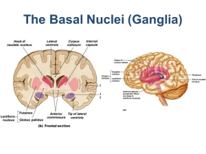
These are 5 masses of grey matter in the subcortical part of the cerebral hemisphere on each side of the brain. They include Caudate nucleus, Lentiform nucleus. The caudate and lentiform together are called corpus striatum, Amygdaloid body, Claustrum, The basal ganglia also include substantia nigra (SN) and subthalamic nucleus.
The caudate nucleus
the caudate nucleus is a curved nucleus formed of head, body, and tail. It is related to the lateral ventricle as follows:
- The head forms the floor & the lateral wall of the anterior horn of the lateral ventricle.
- The body is present in the floor of the central part (body) of the lateral ventricle, This part of the caudate nucleus is regarded as suprathalamic.
- The tail is present in the roof of the inferior horn of the lateral ventricle.
- Antero-inferior, the head of caudate fuses with the lateral part of the lentiform nucleus (putamen).
- Above the site of fusion, the head of caudate is connected with the putamen by strands of grey matter across the anterior limb of the internal capsule, which gives a striated appearance, hence the name corpus striatum was given to the caudate and lentiform.
Lentiform nucleus
It is so-called because it resembles a biconvex lens. It is divided by a band of the white lamina (lateral medullary lamina) into putamen laterally and globus pallidus (GP) medially. The globus is subdivided into medial and lateral segments by another curved lamina (medial or internal medullary lamina) of white matter. It is related medially to the internal capsule and laterally to the external capsule, claustrum, extreme capsule, and insula.
Claustrum
It is a thin sheet of grey matter between the lentiform nucleus and insula; being separated from the lentiform nucleus by the external capsule. The claustrum has afferent and efferent connections with the cerebral cortex. It has to be emphasized that the claustrum has widespread reciprocal connections with the sensory cortex.
Amygdaloid body (Amygdala)
It is an irregular complex of nuclei that lies deep to the uncus it is related to the anterior part of the roof of the inferior horn of the lateral ventricle. It is connected with the olfactory structures and forms part of the limbic system. Its cell axons are named stria terminalis.
Physiology of the basal ganglia
The basal ganglia are five subcortical nuclei on each side of the brain. The globus pallidus is divided into external (GPext) and internal segments (GPint), The caudate and putamen are regarded as the receiving center of the basal ganglia, while the globus pallidus internal segment is regarded as the discharging center, The substantia nigra is divided into 2 parts, the pars compact, and the pars reticulata.
Internal projections of the basal ganglia
There are many internal basal ganglia projections, they include:
- Striato-Pallidal projection; which starts from the caudate and putamen to the globus pallidus. It is inhibitory and utilizes GABA as its transmitter.
- Nigro-Striatal projection: passes from the substantia nigra pars compacta to the corpus striatum, the transmitter is dopamine and it is disordered in Parkinsonism.
- The globus pallidus internal segment projects to the thalamus, using GABA as the transmitter.
Motor pathways of the basal ganglia
The basal ganglia function not by themselves but always in close association with the cerebral cortex of the same side which controls muscles of the opposite side of the body, two closed circuits process signals in the basal ganglia, the direct and the indirect pathways, They start from the cerebral cortex then project to the basal ganglia and back to the cerebral cortex.
1. The direct pathway
The direct pathway starts with cortical excitation of the striatum, When the direct pathway striatal neurons fire in response they release GABA, which inhibits the activity of the GPint neurons, This inhibition releases the thalamic neurons from inhibition dis-inhibit the thalamic neurons), allowing them to fire to excite the cortex, Therefore, the net result of exciting the direct pathway striatal neurons is to excite motor cortex, Dopamine acting on D1 receptors enhances the activity of this circuit.
2. The indirect pathway
The indirect pathway starts with a different set of cells in the corpus striatum. When the indirect pathway striatal neurons are active they release GABA, which inhibits the GPext neurons, This inhibition releases the subthalamic neurons (dis-inhibits the subthalamic neurons), With the subthalamic neurons free to fire glutamate to excite the GPint.
The GPint neurons inhibit the thalamus thereby producing a net inhibition on the motor cortex to suppress inappropriate motor programs, The normal functioning of the basal ganglia involves a proper balance between the activity of these two pathways; stimulation of dopamine (D2) receptors hyperpolarizes and inhibits the indirect pathway striatal neurons.
The nigro-striatal pathway is an important pathway in the modulation of the direct and indirect pathways, This pathway thus has the dual effect of exciting the direct pathway while simultaneously inhibiting the indirect pathway, Because of this dual effect, excitation of the nigro-striatal pathway has the net effect of exciting the cortex.
Functions of the basal ganglia
The basal ganglia participate in 4 pathways: motor, oculomotor, cognitive, and limbic.
1. Motor functions:
- Neurons in the basal ganglia are involved in the planning of movement as they discharge before movements begin.
- The basal ganglia facilitate the ongoing intended voluntary movement through the direct circuit.
- They inhibit and suppress competing or unwanted motor activity through the indirect circuit.
- The caudate and putamen share in learning and storing automatic subconscious movements.
2. Initiation of conjugate eye movements: Eye movements are initiated in the frontal eye field area; however, initiation of eye movements requires the activation of the superior colliculus neurons (which are normally tonically inhibited by substantia nigra pars reticulata neurons), Momentarily inhibition of substantia nigra through the basal ganglia oculomotor pathway, allows activation of the collicular neurons, allowing the change of gaze to a new location.
3. Cognitive function: The basal ganglia are connected to the prefrontal cortex, Through these loops, the basal ganglia are thought to play a role in problem-solving and decision-making organizing behaviors.
4. Emotional function: mediating empathy towards others and motivational behavior.
Disorders of the basal ganglia motor circuits
The loss of the dopamine-containing neurons of the pars compacta of the substantia nigra:
- The loss of these neurons leads to reduced quantities of dopamine reaching the corpus striatum, It results in a decrease in the activity of the direct pathway and an increase activity in the indirect pathway.
- Thus, the GPint neurons are abnormally active, keeping the thalamic neurons inhibited, Without the thalamic input, the motor cortex neurons are not as excited, and therefore the motor system is less able to execute the motor plans in response to the patient’s movement.
- Parkinson’s disease is one of the basal ganglia disorders that result from a decrease in dopaminergic transmission in the nigro-striatal pathway of the basal ganglia. It is a progressive neurological disorder that presents with problems of movement followed by cognitive deficits.
Manifestations of Parkinson’s disease
1. Slowness of movement (bradykinesia): the disease is characterized by slowing of normal movement, and difficulty in initiating movements which are manifested as:
- Reduced blinking.
- Shuffling gait (short stepping gait).
- The affection of various daily simple or complex tasks (e.g. washing, brushing the teeth, dressing, and writing).
- Poorly articulated speech which becomes slow monotonous and low in volume.
2. Resting tremors: coarse static tremors that are seen at rest. They usually begin in one extremity and spread to the others, affecting the distal joints of the limbs preferentially. In the hand, the tremors may involve the thumb and fingers rolling together, so it is called pill-rolling in character.
3. Rigidity:
- There is a marked increase in muscle tone in both flexor and extensor muscles.
- Passive movement of the limb will show either rigidity that is present all through the movement and is called lead pipe rigidity (resistance like bending a lead pipe); or rigidity that can be interrupted by tremors and is called cogwheel rigidity.
- The rigidity results in stiffness and tension in the muscles, which can make it difficult to move around and make facial expressions (expressionless face).
You can follow Science Online on YouTube from this link: Science online
White matter of the brain function, structure and Blood supply of the internal capsule
Function and Physiology of Thalamus, Hypothalamus and Limbic System
Diencephalon function, Thalamus, Metathalamus, Hypothalamus, Epithalamus and Subthalamus
Reticular formation, Reticular activating system and Types of EEG waves and Phases of sleep
Upper and lower motor neurons lesion, Stages of complete spinal cord transection


