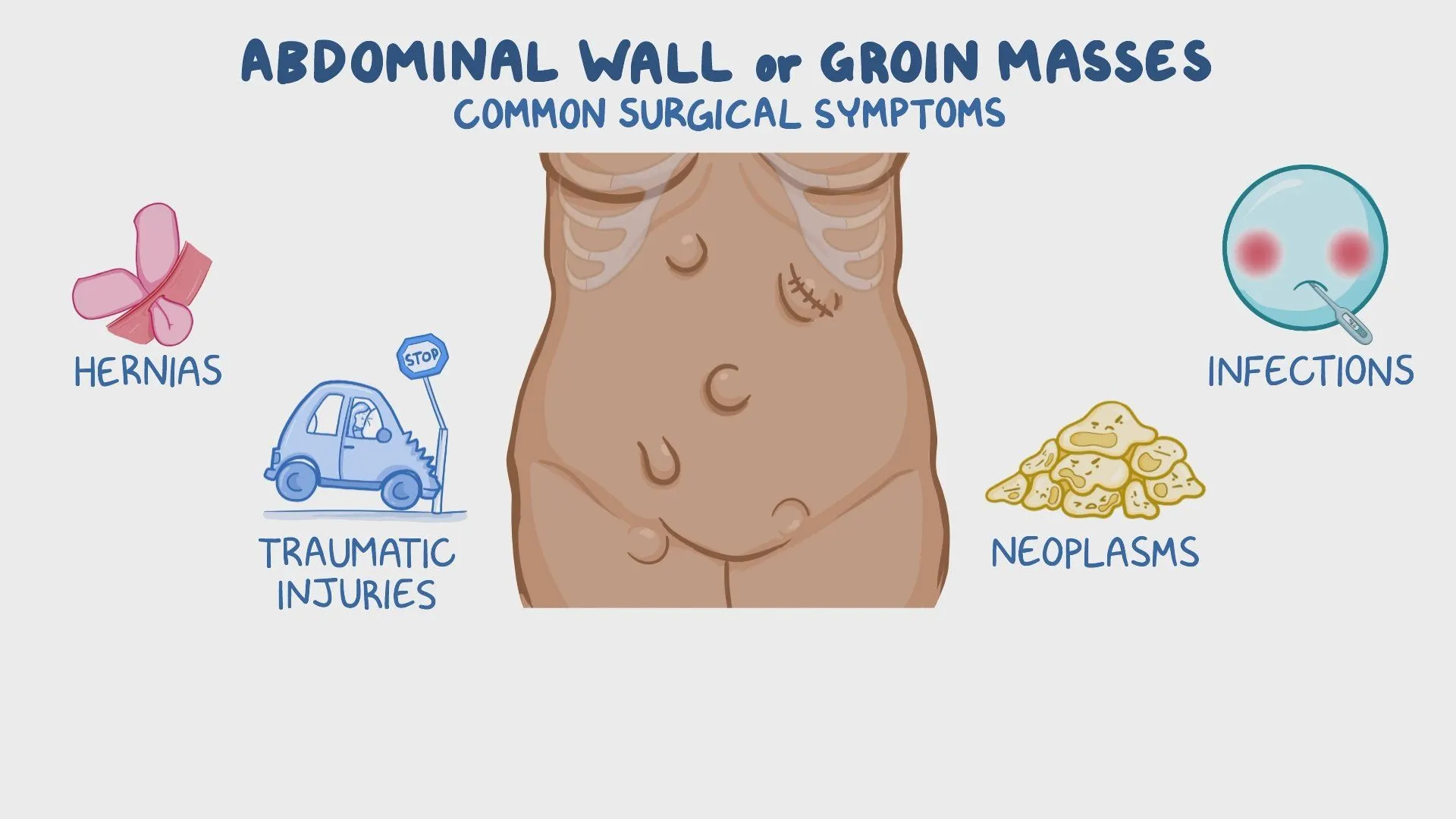
Abdominal masses Symptoms and Treatment, What is the most common abdominal wall mass?
An abdominal mass is an abnormal growth or lump that can be felt in the abdomen. It may be caused by various conditions, ranging from benign (harmless) to malignant (cancerous). The location, size, consistency, and associated symptoms help determine the possible cause.
Symptoms Associated with Abdominal Masses
- Pain or discomfort in the abdomen.
- Bloating or a feeling of fullness.
- Unintended weight loss.
- Nausea, vomiting, or changes in bowel habits.
- Fever (if infection-related).
- Jaundice (if liver or bile duct involvement).
Diagnosis
Physical examination – To assess size, location, and consistency of the mass.
Imaging tests:
- Ultrasound – First-line imaging for abdominal masses.
- CT scan or MRI – Provides detailed views of organs and tumors.
- Blood tests – To check for infection, liver/kidney function, or tumor markers.
- Biopsy – May be needed to confirm the nature of the mass.
Abdominal masses
Abdominal masses
Parietal Swellings:
- Extends over the costal margin.
- Moves ant. & post with respiration.
- More prominent on rising-up test.
Intra-abdominal Swellings
- Disappears beneath costal margin.
- Moves up and down with respiration.
- Less prominent on rising-up test.
Imaging by C.T. to confirm diagnosis (Ultrasound not sufficient).
Parietal Swellings
1. Skin:
- Sebaceous cyst.
- Papilloma.
- Melanoma.
- Squamous cell carcinoma (SCC).
2. Subcutaneous Tissue:
- Lipoma (Subcutaneous or inter-muscular).
- Neurofibroma.
- Hemangioma and Lymphangioma.
3. Muscles:
- Rectus sheath hematoma
- Tumor demolding:
- Recurrent Paget’s fibroma.
- locally malignant tumor.
- Uncapsulated fibroma that arises from the musculoaponrurotic structures of the anterior abdominal wall, below the umbilicus.
Pathology of desmoid tumor: fibrous tissue, epithiliod cells “multinucleated”.
Etiology of desmoid tumor:
- may be related to trauma.
- On top of surgical scars (common in the site of scar of caesarean section).
- stretch of muscles fibers e.g.” in pregnant woman “
- Gardner’s syndrome (FAP).
Clinical picture of desmoid tumor:
- more in female.
- site: below umbilicus.
- Swelling: hard in consistency and mobile from side to side.
Treatment: Complete excision with a safety margin to prevent recurrence.
Intra-abdominal Swellings
1. Right hypochondrium masses:
- hepatomegaly
- Enlarged lost bladder.
- Mass in upper pole of right kidney or suprarenal gland.
- Cancer in hepatic flexure of colon
2. Right Latin masses:
- Mass in right kidney or suprarenal gland.
- Mass in ascending Colon.
- Retroperitoneal masses Lymphoma, teratoma).
3. right iliac fossa mass
- Carcinoma of the cecum.
- Appendicular mass or abscess.
- ileocaecal tuberculosis.
- Amoeboma.
- Psoas abscess.
- Bony masses.
- Ovarian/uterine masses.
- LN mass: mesenteric or external Iliac LNs.
- Undescended testis.
- Crohn’s disease.
- Ectopic (pelvic) kidney.
4. Epigastric masses
- Left lobe of the liver.
- Gastric tumors.
- Pyloric stenosis.
- Duodenal masses.
- Pancreatic mass.
- Transverse colon cancer
- Para-aortic LN mass.
- Abdominal Aortic Aneurysm.
- Retroperitoneal masses (Lymphoma, teratoma).
5. Central abdominal (umbilical) mass
- Mesenteric mass or cyst.
- Omental cyst.
- Ovarian cyst (pedunculated).
- Small bowel tumours.
- Transverse colon mass.
- Mass in body of pancreas.
- Retroperitoneal masses.
- LNs mass
- Aortic aneurysm
6. Supra-pubic mass
- enlarged bladder
- uterine mass e.g. fibroids.
- pregnant uterus.
- Ovarian mass e.g. cyst.
7. Left hypochondrium masses
- Splenomegaly.
- Mass arising from the tail of the Pancreas.
- Mass in upper pole of left kidney or suprarenal gland.
- Cancer in splenic flexure of colon.
8. Left loin masses
- Mass in left kidney or suprarenal gland.
- Mass in descending Colon
- Retroperitoneal masses (Lymphoma, teratoma).
Left iliac fossa mass
- Carcinoma sigmoid or descending colon.
- Psoas abscess.
- Bony masses.
- Ovarian/uterine masses
- LN mass: mesenteric or external Iliac LNs.
- Undescended testis.
- Crohn’s disease.
- Ectopic (pelvic) kidney.
Common Causes of Abdominal Masses
Gastrointestinal Causes:
- Hernia – A bulge due to the protrusion of intestines through the abdominal wall.
- Colon cancer – Can present as a palpable mass in advanced stages.
- Intestinal obstruction – A blockage that may cause a distended abdomen.
Hepatobiliary Causes (Liver & Gallbladder):
- Hepatomegaly – Enlarged liver due to cirrhosis, hepatitis, or cancer.
- Liver tumors (benign or malignant, e.g., hepatocellular carcinoma).
- Gallbladder enlargement (cholecystitis, gallbladder cancer).
Pancreatic Causes:
- Pancreatic cysts or tumors – Can present as an upper abdominal mass.
- Pancreatitis (pseudocyst formation).
Renal (Kidney) Causes:
- Polycystic kidney disease (PKD) – Enlarged kidneys due to multiple cysts.
- Kidney tumors (renal cell carcinoma).
- Hydronephrosis – Swelling of the kidney due to urine buildup.
Gynecological Causes (in women):
- Ovarian cysts or tumors – May present as a lower abdominal mass.
- Uterine fibroids – Benign growths in the uterus.
- Pregnancy-related masses (ectopic pregnancy, molar pregnancy).
Lymphatic Causes:
- Lymphoma – Cancer affecting lymph nodes in the abdomen.
- Lymphadenopathy (enlarged lymph nodes due to infection or malignancy).
Soft Tissue & Miscellaneous Causes:
- Lipoma – A benign fatty tumor.
- Abscess – Infection leading to a pus-filled mass.
- Retroperitoneal tumors – Growths behind the abdominal cavity.
Treatment
Treatment depends on the cause of the abdominal mass:
- Surgical removal – For tumors, hernias, or large cysts.
- Medications – Antibiotics for infections, chemotherapy for cancers.
- Drainage procedures – For abscesses or fluid-filled masses.
You can subscribe to Science Online on YouTube from this link: Science Online
Gastroesophageal Reflux Disease, Complications of GERD and Barrett’s oesophagus
Esophagus diseases, Dysphagia causes, Achalasia, and Symptomatic Diffuse Esophageal spasm
Pharynx function, anatomy, location, muscles, structure, and Esophagus parts
Tongue function, anatomy, and structure, Types of lingual papillae, and Types of cells in taste bud
Mouth Cavity divisions, anatomy, function, muscles, Contents of Soft palate and Hard palate
Temporal and infratemporal fossae contents, Muscles of mastication and Otic ganglion