Structure of leaf and green plastid, Mechanism of photosynthesis in green plants
Green leaves are the main sites of photosynthesis as they contain the green plastids in the higher plants, Green herbaceous stems may participate, to some extent, in this process as they contain chlorenchymatous tissues which contain green plastids.
Structure of green plastid
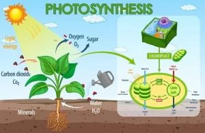
Green plastid (Chloroplast) appears under the light microscope as a homogeneous mass, having the shape of a convex lens, After studying the green plastid under the electron microscope, it was found that it consists of a Double thin external membrane, Matrix (Stroma), Starch granules and Grana.
Green plastid consists of a double thin external membrane and its thickness is about 10 nm, Matrix (Stroma) is a colourless protein substance.
Starch granules are large in number, They are small in size and a temporary product of the photosynthesis process as they will soon change back into a soluble sugar to be translocated under certain conditions to other organs of the plant.
Grana are located in the stroma, They have disc-shaped structures, each granum is about 0.5 micron in diameter and about 0.7 micron in thickness, Each granum is a pile (mass) of 15 hollow discs or more that are arranged above each other.
The margin of some discs of a granum extends to meet the margin of another disc in the neighbouring granum to increase the surface area of grana that are exposed to the light, They contain the pigments which absorb the light energy needed for photosynthesis.
Main four pigments in chloroplates:
- Chlorophyll A: Its colour is blue-green, the ratio is 70 %.
- Chlorophyll B: Its colour is Yellow-green, the ratio is 70 %.
- Xanthophyll: Lemon-yellow, ratio is 25 %.
- Carotene: Orange-yellow, ratio is 5 %.
The green colour dominates over the other colours of pigments due to the high ratio of the chlorophyll pigments.
The importance of chlorophyll is the absorption of light energy needed for the photosynthesis process, The structure of chlorophyll is a complicated structure and the molecular formula of chlorophyll ( A ) is C55H72O5N4Mg.
It is believed that there is a relation between the presence of the Mg atom in the centre of the chlorophyll molecule and the ability of chlorophyll to absorb light.
Structure of the leaf
Examination of T.S. passing through the midrib of a dicotyledonous plant leaf, the leaf consists of three main tissues which are upper and lower epidermis, Mesophyll tissue, and vascular tissue.
Upper and lower epidermis
Each of the upper and lower epidermis consists of one row of adjacent barrel-shaped parenchyma cells with no chlorophyll, Stomata spread throughout the epidermal layer, and the external wall of the epidermis is coated by a layer of cutin, except the stomata.
Mesophyll tissue
It lies between the upper and lower epidermal layers and is transversed by veins, It consists of two layers which are the palisade layer and the spongy layer.
The palisade layer is perpendicular to the upper epidermis of the leaf surface, It consists of one row of cylindrical and elongated parenchyma cells, It possesses many chloroplasts (especially in the upper part of cells) to receive the highest light intensity.
The spongy layer is located below the palisade tissue, It consists of irregular-shaped and loosely-arranged parenchyma cells with wide intercellular spaces, The cells of this tissue contain a lower number of chloroplasts than the cells of palisade tissue.
Vascular tissue
It consists of numerous vascular bundles that extend through the veins and venules, The midrib contains the main vascular bundle of the leaf, The vascular bundle consists of Xylem vessels and Pheloem.
Xylem vessels: several vertical rows that are separated by thin-walled cells called xylem parenchyma.
Phloem: It lies toward the lower epidermis, It translocates the dissolved organic food from the mesophyll where it has been manufactured to the other parts of the plant.
Mechanism of photosynthesis
Source of oxygen evolved during photosynthesis: The American scientist Van Neil was the first person who point out the source of oxygen evolved in photosynthesis by studying this process in the green and purple sulphur bacteria.
Green and purple sulphur bacteria
Green and purple sulphur bacteria are autotrophic as they contain bacteriochlorophyll which is simpler in structure than the normal chlorophyll.
They live in swamps and ponds, because hydrogen sulphide is abundant which is the source of hydrogen used by these bacteria to reduce CO2 to build up carbohydrates and sulphur is released.
Van Neil assumed that: Light decomposed hydrogen sulphide into hydrogen and sulphur.
12H2S → 12H2 + 12 S ↓
The resulted hydrogen was used in certain dark reactions to reduce CO2 into carbohydrates.
12H2 + 6 CO2→ C6H12O6 + 6 H2O ↓
So, the general equation for photosynthesis in sulphur bacteria is:
6 CO2 + 12H2S→ C6H12O6 + 6 H2O + 12 S ↓
Green plants
Van Neil assumed that:
Light decomposed water into hydrogen and oxygen.
12H2O → 12H2 + 6 O2 ↑
The resulted hydrogen was used in certain dark reactions to reduce CO2 into carbohydrates.
12H2 + 6 CO2 → C6H12O6 + 6 H2O
So, the general equation for photosynthesis in green plants is:
6 CO2 + 12H2S→ C6H12O6 + 6 H2O + 6 O2 ↑
Consequently, Van Neil proposed that: the release of oxygen comes from water in the green plants, a case which is similar to sulphur that is released from hydrogen sulphide by the sulphur bacteria.
To confirm that water is the source of oxygen gas that evolved from the photosynthesis process, In 1941, a group of scientists at California University verified the theory of Van experimentally by using a green agla called Chlorella and provided it with all conditions favourable for photosynthesis, The result: The source of evolved oxygen from photosynthesis is water and not CO2 gas.
Asexual and sexual reproduction in plants, Pollination and Stages of fertilization process in plants
Autotrophic Nutrition in green plants, Mechanism of water and minerals absorption


