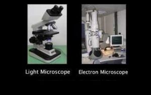
Light microscope and Electron microscope
It is difficult to see the cell or its components due to their very minute size , So, the cell discovery was related to the invention of the microscope , Also, seeing of cell components was related to the interesting of its magnifying power .
The studies of the cell structure were crowned with the invention of the electron microscope which has a high magnifying power, So, There are two types of the microscopes which are the light microscope and the Electron microscope.
Light microscope
Until 1950 , the light microscope was the only device available for scientists to examine the micro-organisms , It depends on the sunlight or an artificial light to work .
Functions of the light microscope:
- Magnifying many micro-organisms and non-living things .
- Examining large-sized objects after cutting them into very thin slices that allow the light to transmit through them.
Magnifying power magnifies the objects up to 1500 times its real size , Magnification depends on the magnifying power of its ocular and objective lenses, Light microscope can not magnify the objects more than 1500 times because the image becomes unclear ( blurred ) .
Microscope magnifying power = the magnifying power of ocular lens × the magnifying power of the objective lens
Methods of obtaining most clear image by using light microscope :
Scientists found that the best method to examine the specimens more clearly is the increasing of contrast between different parts of the specimens by :
- Using dyes to stain or colour certain parts of the specimen to be more clearer but these dyes kill the living specimens ( disadvantages ) .
- Changing the level of light .
The compound light microscope is used to magnify and examine the tiny things and their contents .
Electron microscope
In 1950, scientists started to use the electron microscope , Idea of its work depends on using a beam of high speed electrons instead of the light, These electrons are controlled by the electromagnetic lenses , Magnifying power magnifies the objects one million times of their real size . .
Functions of the electron microscope:
- Clarifying cellular components that had not been known before.
- Knowing more accurate details for the structure that had been known before .
Properties of its images ( micrograghs ) :
- They are high resolution magnified and highly contrasted comparatively to those produced by the light microscopes due to the shortness of wavelength of the electronic ray comparatively to the light ray .
- They are received on a highly sensitive fluorescent screen or on a photographing board .
Types of the electron microscope
- Scanning electron microscope that is used to study the cell surface.
- Transmission electron microscope that is used to study the cell internal structure.
- The micrograph of the white blood cell is more clearer by using transmission electron microscope due to the easiness of distinguishing its internal components.
You can download the application on google play from this link: Science online Apps on Google play
Compton effect, Photon properties, Electron microscope and Optical microscope


