Human Transport system, Structure of human circulatory system (heart, blood vessels & blood)
Animals obtain their requirements of energy in the form of food, after its digestion, the soluble food substances are absorbed, but still remains the problem of their transport and distribution to the various tissues laying far away from the surface of absorption.
In small animals ( Protozoa and Hydra ): Both gases & food substances are transported by diffusion, In bigger and more complicated animals: Diffusion is not sufficient for the transportation of food and oxygen to the various tissues, Therefore, the presence of a specialized transport system is essential in these animals.
Transport in human
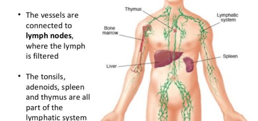
The process of transport takes place in the human body through two systems that are closely connected with each other, these systems are the Blood vascular system ( Circulatory system ) and the Lymphatic system.
Circulatory system
The circulatory system in human is considered from the closed type, as the blood vessels are connected together with the heart in a complete circuit through which the blood passes, Structure of human circulatory system: Heart, blood vessels, and blood.
Heart
The heart is a hollow muscular organ that lies nearly in the middle of the chest cavity, It is enclosed in the pericardium, to protect the heart and facilitate its action, The heart beats regularly throughout the whole lifetime.
The heart is divided horizontally into four chambers: The upper two thin-walled chambers are called atria which receive the blood from the veins, The lower chambers with thick muscular walls are called ventricles that pump the blood through the arteries.
The heart is divided longitudinally into two sides using muscular walls which are the right side and left side, Each side has an atrium and a ventricle connected together by an opening that is guarded by a valve with thin flaps whose free edges are attracted by tendons to the ventricle wall, to prevent the flaps from turning inside out, Thus, the blood is permitted to flow from the atrium to the ventricle, but not in the reverse direction.
Types of heart valves
- The tricuspid valve ( consists of 3 flaps ) is between the right atrium and right ventricle, It allows the blood to pass from the right atrium to the right ventricle (in one direction), but not in the reverse direction.
- The bicuspid valve (Mitral valve) (consists of 2 flaps) is between the left atrium and left ventricle, It allows the blood to pass from the left atrium to the left ventricle (in one direction), but not in the reverse direction.
- Semilunar valves (Aortic valve and pulmonary valve) are at the connection of the heart with both the aorta and pulmonary artery, They allow the blood to pass from the two ventricles to the arteries in one direction (prevent the blood from returning to the ventricles).
Heartbeats: The rhythmic heartbeats are spontaneous, as they originate from the cardiac tissue itself because it has been proven that the heart continues beating regularly even after it has been disconnected from the body and cardiac nerves, The rhythmic heartbeat is caused due to the presence of two muscular nodes, which are:
Sino-atrial node
It is a specialized bundle of thin cardiac muscular fibers that is buried in the right atrial wall near the connection between the right atrium (auricle) and the large veins, It is considered as the pacemaker of the heart, as it beats at a regular rate of 70 beats/minute (normal rate).
It is connected to two nerves: A nerve decreases the rate of heartbeats during sleeping and in sadness states (grief), and a nerve increases the rate of heartbeats during joy, after waking up, and in case of severe physical effort, So, The number of cardiac beats changes according to the physical and psychological states of the body.
Atrio-ventricular node
Atrio-ventricular node is located at the junction between the atria and ventricles, Mechanism of heartbeats:
- The sino-atrial node sends impulses over the two atria which are stimulated to contract.
- The electrical impulses reach the atrio-ventricular node.
- The impulses will spread rapidly through special fibers (His fiber) from the inner-ventricular septum to the wall of both ventricles, where they stimulate their muscles to contract.
We can distinguish two sounds of heartbeats :
- Long and low-pitched (Lubb): Due to the closure of two valves between the atria and ventricles during the ventricular contractions.
- Short and high-pitched (Dupp): Due to the closure of aortic and pulmonary valves during the ventricular relaxation.
In the normal age of a man, the heart beats by a range of 70 beats/minute and pumps 5 liters of blood in each minute which is equal to the total amount of blood in the body.
Blood vessels
They include arteries, veins, and blood capillaries.
Arteries
They are vessels that carry the blood from the heart to the other body organs, They are usually buried among the body muscles, All of them carry oxygenated blood, except the pulmonary artery which comes out from the right ventricle to the lungs carrying deoxygenated blood.
The wall of an artery is built up essentially of three layers of tissues, which are:
- The outer layer consists of a coat of connective tissue.
- The middle layer is relatively thick and consists of involuntary muscles that contract and relax under the control of nerve fibers, thus it can pulsate to pump blood.
- The inner layer ( Endothelium ) consists of one row of tiny epithelial cells that are provided with elastic fibers that give elasticity to the artery to be able to pump blood during the contraction of ventricles.
Veins
They are vessels that carry blood from all the body parts to the heart, They carry deoxygenated blood, except the pulmonary veins which open into the left atrium (autricle) carrying oxygenated blood.
The wall of the vein, like the artery, is composed of the same three layers, but with certain modifications, which are less elastic fibers ( rare ), The middle layer is less thick, so, the wall of the vein is thinner than that of the artery and does not pulsate.
Several veins possess a system of internal valves along their length to prevent the back-flow of blood, allowing only the passage of blood in the direction of the heart.
Sites of these valves can be observed in the arm veins when the arm is tied tightly with a bandage (tourniquet) above the elbow, this was done by the English doctor William Harvey, who described blood circulation in the 17th century, William Harvey studied the blood circulation in the 17th century after being discovered by the Arab doctor Ibn Al-Nafis in the 10th century.
Capillaries
They are tiny microscope vessels that connect the arterioles with the venules, This fact was discovered by the Italian scientist Malpighi at the end of the 17th century, and thus completed the work of Harvey.
Capillaries spread in the spaces between cells all over the body tissues because they reach all the cells and supply them with their requirements of food and oxygen.
The wall is About 0.01 micron (0.00001 mm) thick, which facilitates the quick exchange of substances between the blood and tissue cells, It consists of one row of thin epithelial cells with tiny pores between them, The diameter is the average diameter of (7 – 10 micron).
Heart function, structure, Valves, Borders, Chambers and Surfaces
Blood pressure, structure, functions and Mechanism of blood clotting


