Function of Cytoplasmic organelles, Mitochondria, Peroxisomes & Cytoskeleton
The cytoplasm is all of the materials within the cell, It is enclosed by the cell membrane, except for the cell nucleus. The organelles within the cytoplasm do not contain any of a cell’s genetic material, because all of that is exclusively contained within the nucleus. The organelles inside the cytoplasm are very important for the maintenance of the cell.
Cytoplasmic organelles
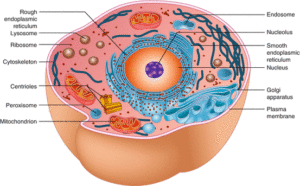
The cytoplasm is the substance of the cell other than the nucleus and cell membrane, and it is basically fluid in nature, “Organelles” are the functional elements within the cytoplasm. Some of the most important organelles that cytoplasm contains are the ribosomes, mitochondria, proteins, the endoplasmic reticulum, lysosomes, and the Golgi apparatus.
Mitochondria
The mitochondria are powerhouses of the cell as they are the sites of adenosine triphosphate (ATP) production.
LM picture
Mitochondria, when present in large numbers contribute to cytoplasmic eosinophilia because of the large amount of membrane they contain. Mitochondria can be visualized in tissues by using special stains e.g. silver stain.
EM picture
Mitochondria are membrane-bounded organelles, present in all types of cells except in RBCs and terminal keratinocytes of skin epidermis. They have variable shapes and their number ranges from few to several hundred depending on the energy needs of the various cell types. Each mitochondrion is surrounded by two membranes: outer and inner, which define two mitochondrial compartments;
- The intermembranous space, the compartment located between the two membranes.
- The matrix space, the compartment enclosed by the inner membrane.
Mitochondrial membranes contain distinct protein composition related to its function, The outer membrane is smooth and porous. It allows easy passage of small molecules due to the presence of specific transmembrane proteins called mitochondrial porins.
The inner membrane
- It is folded into numerous cristae which greatly increase its total surface area, the number of cristae is greater in cells of greater demand for ATP as in cardiac muscle fibers.
- Most mitochondria have flat, lamellar cristae in their interiors, whereas cells that secrete steroids frequently contain tubular cristae.
- It is highly impermeable to ions and small molecules due to the presence of specific phospholipid called cardiolipin.
- It contains the enzymatic components of the electron transport system (respiratory chain enzymes) and the ATP synthase. These enzymes from the elementary particles projecting from the mitochondrial cristal membrane into the matrix.
Matrix space is surrounded by the inner mitochondrial membrane. It contains the following:
- Numerous soluble enzymes involved in specialized mitochondrial functions as a citric acid cycle.
- Mitochondrial DNA and few ribosomes.
- Matrix granules that store calcium ions (Ca+) thus play a role in mitochondrial regulation of Ca+ intracellular concentration.
Intermembranous space contains substrates diffusing from the cytoplasm through the outer membrane and ions pumped out of the matrix space through the inner membrane.
The genetic system of mitochondria: Mitochondria are self-replicating organelles, the mitochondrial genome is a circular DNA molecule with limited coding capacity. It represents 1% of the total DNA of the cell. Mitochondria can synthesize some of their structural proteins by their own RNAs. Most of the mitochondrial proteins are encoded by the nuclear DNA and are synthesized in the cytoplasm and subsequently imported into mitochondria.
Peroxisomes
Peroxisomes are membrane-bounded organelles that contain oxidative enzymes involved in the oxidation of several substrates. Peroxisomes possess no genetic material of their own.
LM picture
They are not seen by H & E stain.
EM picture
They are small, spherical microbodies with fine granular electron-dense content bounded by a single membrane.
Functions of peroxisomes
- β-oxidation of long-chain fatty acids to generate energy. However, they differ from mitochondria in that they are unable to store this energy in the form of ATP. This energy is released as heat and contributes to the maintenance of body temperature.
- Generation of hydrogen peroxide, which detoxifies various toxic agents.
- Contain catalase enzyme that converts the excess hydrogen peroxide, which is toxic, into the water, thus protecting the cell.
- Detoxification of alcohol in cooperation with the smooth endoplasmic reticulum in the liver.
Cytoskeleton
The cytoskeleton is a network of structural proteins belonging to the non-membranous cell organelles. There are 3 components of cytoskeleton depending on their thickness and their structural proteins, these are the microfilaments, microtubules, and intermediate filaments.
Microfilaments (actin filaments)
- Diameter: about 7nm.
- LM picture: can be visualized by using immunohistochemical staining.
- EM picture: they appear as thin electron-dense filaments.
- Structural proteins: an actin filament consists of monomers of G-actin (globular actin) polymerize to form F-actin (filamentous actin) arranged as a double helix.
Function: microfilaments are dynamic structures that can elongate and shorten thus, they participate in:
- Cell motility for cell migration, Cytoplasmic streaming during movement of organelles and intracellular transport of vesicles within the cytoplasm, Cytokinesis; formation of contractile ring during cell division, and Muscle contraction associated with the motor protein, myosin.
- Structured role in the maintenance of the shape of the cell, Formation the structured core of microvilli.
Microtubules
- Diameter: about 25 nm.
- LM picture: can be visualized by using immunohistochemical staining.
- EM picture: they appear as fine tubules.
- Structured proteins: It consists of a globular protein dimer called tubulin (each is composed of alpha and beta subunits). Chains of tubulin dimers form a protofilament. The wall of a microtubule is made up of 13 protofilaments that run longitudinally. motor proteins associated with microtubules.
Function: microtubules, like microfilaments, are dynamic structures that can elongate and shorten thus, they participate with actin in:
- Cell motility for the transport of organelles and vesicles in the cytoplasm.
- Structured role in the formation of the mitotic spindle to separate the chromosomes during cell division, and the formation of centrioles, cilia, and flagella.
Intermediate filaments
- diameter: about 10 nm.
- LM picture: intermediate filaments can be visualized by using immunohistochemical staining.
- EM picture: they appear as electron-dense filaments thicker than actin filaments.
- Structural proteins: Intermediate filaments are constructed like woven ropes.
Function and classification: Intermediate filaments are the most stable and permanent members of the cytoskeletal elements thus they play a structural role and do not participate in cell motility. They are classified according to their protein composition and their specific cellular distribution into the following:
- Keratin (cytokeratin) filaments: they are found in epithelial cells.
- Vimentin found in the cells of mesenchymal origin e.g. fibroblasts.
- Desmin in muscle cells.
- Glial fibrillary acidic protein in neuroglial cells e.g. astrocytes.
- Neurofilaments in nerve cells. These 5 types of intermediate filaments are cytoplasmic i.e. located in the cytoplasm.
- Nuclear lamins are the nuclear intermediate filaments that form a network lining the inner side of the nuclear envelope forming the nucleoskeleton (nuclear lamina).
Clinical importance of intermediate filaments
Hence the intermediate filaments (except for the lamins) are located in specific tissue types, they can be used to determine the histogenesis of tumors. Immunohistochemical staining of each type of the intermediate filaments allows the recognition of the primary origin of a malignant tumor.
Cytoplasmic organelles, Ribosomes & Endoplasmic reticulum function, structure & definition
Cell Structure, the function of Golgi apparatus, Endosomes & Lysosomes
Nucleus components, function, diagram & classification of chromosomes
Histology, Molecular structure of the cell membrane, Cell function & structure


