Physiology of central human reflexes, Types and properties of Spinal cord reflexes
In biology, The reflex, or reflex action, is an involuntary, unplanned sequence or action and nearly instantaneous movement in response to a stimulus, The reflex is made possible by neural pathways called reflex arcs which can act on an impulse before that impulse reaches the brain, The reflex is an automatic response to a stimulus that does not receive or need conscious thought.
Physiology of central human reflexes
A reflex is defined as the involuntary response of an organ to a stimulus. There are several ways of classifications:
According to the center:
- Hypothalamic reflexes.
- Midbrain (mesencephalic) reflexes.
- Medullary reflexes.
- Spinal reflexes.
According to the location of receptors:
- Superficial reflexes; result from the stimulation of skin receptors.
- Deep reflexes; result from the stimulation of muscle and tendon receptors.
- Visceral reflexes; result from the stimulation of visceral receptors as micturition and defecation reflexes.
According to the number of synapses:
- Monosynaptic reflexes; the center of the reflex is formed of only one synapse.
- Polysynaptic reflexes; the center of the reflex is formed of multiple synapses; if only 2 synapses it is called disynaptic.
Spinal cord reflexes
I. Superficial reflexes
They result from the stimulation of receptors in the skin.
- Plantar reflex: scratching the lateral edge of the sole with a blunt object produces plantar flexion of the toes.
- Flexor withdrawal reflex: application of an injurious stimulus to skin results in contraction of the flexor muscles of the limb withdrawing it away from the stimulus with inhibition of the extensor muscles. It is a protective reflex. The flexor reflex is a typical polysynaptic reflex with a central delay of more than 3 milliseconds. It shows the phenomena of irradiation reciprocal innervation and recruitment with after discharge.
- Crossed extensor reflex: this is the extension of a limb during flexion of the other limb as a result of the withdrawal reflex. It is a supportive reflex.
- Upper and lower abdominal reflexes: striking the abdominal skin by a pin, starting laterally and towards the umbilicus causes contraction of the underlying abdominal muscles. They are a form of withdrawal reflex.
II. Deep reflexes
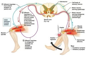
They result from the stimulation of receptors present in muscles and tendons. They include the stretch reflex. This is a monosynaptic reflex elicited by applying stretch to a muscle. The result is a reflex contraction of the stretched muscle. It is the basis of both muscle tone and tendon jerks.
Ill. Visceral (autonomic reflexes)
- The micturition reflex.
- The defecation reflex.
- The erection reflex.
The center of these 3 reflexes is in S 4 & 3,2.
General properties of reflexes
1. Irradiation
The extent of the response in a reflex is directly proportional to the intensity of the stimulus. The more intense the stimulus, the greater is the spread of excitatory impulses up and down the spinal cord to involve more and more motor neurons, e.g. a weak painful stimulus applied to the sole leads to dorsiflexion of the ankle while a stronger stimulus results in flexion of the knee and hip.
2. Forward direction
In a reflex action, impulses pass only from afferent to efferent neurons and never in the opposite direction.
3. Summation
The two types of summation, spatial and temporal, occur in reflex action. Excitation field is the neuronal pool that receives the terminal knobs of a certain afferent nerve. The central part of this field is called the discharge zone because the neurons in this zone receive a large number of synaptic knobs and their excitability is increased to the threshold of discharging.
On the other hand, the peripheral parts of the excitation field are known as the subliminal (or subminimal) fringe, because the neurons in this zone receive a few numbers of synaptic knobs and they are only facilitated (but they do not discharge).
4. Occlusion
Occlusion means, when a muscle is made to contract reflexly, the tension developed by the simultaneous stimulation of 2 near afferents is less than the sum of tensions developed by stimulation of each of the 2 afferents separately. This phenomenon is called occlusion and is due to the overlap of the discharge zones of near afferent fibers.
5. Subliminal fringe
When a muscle is made to contract reflexly, the tension developed by the simultaneous stimulation of 2 afferents is greater than the sum of tensions developed by stimulation of each of the 2 afferents separately. This is due to the overlap of the subliminal fringe zones of both afferent fibers.
6. Reflex delay
This is the time that elapses between the application of the stimulus to the receptor and the appearance of the response. It includes the following:
- The time needed for conduction of impulses along the afferent and efferent fibers.
- The time needed for the conduction of impulses in the nervous system across the synapses, which is the central delay. It is about 0.5 milliseconds per synapse and is an indicator to the number of synapses in a reflex arc.
7. Reflex fatigue and block
If a reflex is repeated successively at short intervals, the response is initially facilitated then becomes progressively weaker. This is due to less liberation of the neurotransmitter in the synapses leading to synaptic fatigue. Fatigue is followed by block i.e. complete stoppage of the response due to depletion of the neurotransmitter stores.
8. Reciprocal innervation
In a reflex action, when a certain group of muscles contracts, the antagonistic group relaxes to the same degree. This occurs via the presence of interneurons that release an inhibitory transmitter at its junction with the AHC supplying the antagonistic muscle.
9. Recruitment and after discharge
In a reflex action, the muscle tension rises gradually due to the gradual activation of the motor neurons. The motor neurons are said to be recruited one after the other. This recruitment of motor neurons is due to different conduction velocities in the afferent fibers and the presence of various interneurons with a variable number of synapses.
While after discharge denotes that, when stimulation is stopped, the muscle relaxes gradually because the motor neurons are still activated by impulses arriving through the reverberating circuits and also caused by the same causes of recruitment.
Reverberating circuits: a type of closed chain interneuron circuit in which interneuron 1 stimulates an output neuron as well as interneuron 2 via a collateral branch, then interneuron 2 stimulates interneuron 1 which re-stimulates the output neuron and interneuron 2 and so on. It is obvious that entry of a brief afferent signal into these circuits causes reverberation (recirculation) of the signal for prolonged periods leading to repetitive excitation of its efferent neurons.
You can follow science online on YouTube from this link: Science online
You can download Science online application on Google Play from this link: Science online Apps on Google Play
Stretch reflex (myotatic reflex) types, properties, Muscle tone, muscle spindles & Clonus
Physiology & function of the spinal cord, Lateral & medial brainstem pathway
Histological organization of spinal cord, Relation between spinal & vertebral segments
Features of synaptic transmission, Mechanism of sensitization & long term potentiation
Synaptic transmission steps, Synapses types & Nature of the postsynaptic change
Blood supply of CNS (central nervous system), Carotid system & Circle of Willis
Skull function, anatomy, structure, views & Criteria of neonatal skull


