Blood supply of CNS (central nervous system), Carotid system and Circle of Willis
The CNS or central nervous system consists of the brain and spinal cord, It is the most important part of the nervous system for maintaining and producing behavior, The brain controls behavior and regulates the body’s physiological processes. It receives information from the body’s sense receptors, which it achieves through communication with the spinal cord, It uses 20 % of the total oxygen we breathe in.
Blood supply of CNS
Arterial supply of the central nervous system:
The arterial supply of the brain comes chiefly from 2 main sources:
- Vertebro basilar system.
- Internal carotid system.
Vertebrobasilar system: it consists of:
- The fourth part of the vertebral artery (two).
- Basilar artery (single).
- Posterior cerebral artery (two).
Vertebral artery (the fourth part)
Origin: the vertebral artery arises in the neck from 1st part of the subclavian artery.
Course & relations: the course of the vertebral artery is divided into 4 parts.
- The 1st part from its origin to the foramen transversarium of the 6th cervical vertebra
- The 2nd part ascends to foramen transversorium of atlas
- The 3rd part lies in the suboccipital triangle.
- The fourth part of the artery enters the skull through the foramen magnum. It ascends on each side of the medulla oblongata in front of the rootlets of the hypoglossal nerve.
Termination: the two vertebral arteries (right and left) unite together at the lower border of the pongs to form a single artery; a basilar artery.
Branches:
- The anterior spinal artery; is a branch from each vertebral artery that unites together to form a single trunk. This trunk runs in the anterior median fissure of the spinal cord.
- Posterior spinal artery, Two arteries, each one arising from the corresponding vertebral artery. Each artery is divided into two longitudinal branches which descend on each side of the posterior root of the spinal nerves.
- The posterior inferior cerebellar artery (PICA) is a tortuous artery and is the largest branch of the vertebral artery
- Medullary branches: to supply the medulla oblongata.
Basilar artery
Origin: at the lower border of pons by the union of the 2 vertebral arteries.
Course & relations: it passes on the front of the pons in the midline (pontine sulcus, groove for basilar artery). The artery is related to the abducent nerve (at its beginning) and oculomotor nerve (at its termination).
Termination: at the upper border of pons by bifurcation into the 2 posterior cerebral arteries.
Branches:
- Anterior inferior cerebellar artery (AICA): supplies the anterior part of the inferior surface of the cerebellum including the cerebellar tonsil.
- Superior cerebellar artery: supplies the superior surface of the cerebellum.
- Pontine branches; supply the pons.
- The Labyrinthine (internal auditory) artery, supplies the inner ear.
- Posterior cerebral artery (terminal branches).
Posterior cerebral artery
Origin: the 2 posterior cerebral arteries begin at the upper border of the pons as the 2 terminal branches of the basilar artery.
Course & relations: the artery passes around the midbrain to reach the occipital pole of the cerebrum. It sinks inside the calcarine sulcus.
Termination: it terminates by dividing into its terminal cortical branches.
Branches:
- Central.
- Cortical:
- Superolateral surface: occipital lobe + lowermost 1 finger breadth of this surface.
- Inferior surface: the whole tentorial surface except temporal pole.
- Medial occipital lobe.
Carotid system
It consists of the intracranial part of the internal carotid artery and its two terminal branches; (the anterior and middle cerebral) arteries.
Internal carotid artery (intracranial part)
The ICA enters the middle cranial fossa by passing through the carotid canal then through the upper part of the foramen lacerum, then it passes Inside the cavernous sinus, lastly, it pierces the roof of the cavernous sinus and passes backward above it. The abducent nerve lies infero-lateral to the ICA. At the base of the brain below the anterior perforated substance, the artery terminates into anterior and middle cerebral arteries.
Anterior cerebral artery
It is smaller at the 2 terminal branches of the ICA. It passes on the medial surface of the brain below the rostrum then in front of the genu then backward above the corpus callosum through the callosal sulcus. It gives cortical branches and central branches.
Middle cerebral artery
It is the larger of the two terminal branches of the ICA. It passes through the stem of the lateral sulcus then in the posterior ramus of the lateral sulcus. It gives central branches and cortical branches.
Cortical branches of the cerebral arteries
The superolateral surface: It is supplied by the middle cerebral artery except: one finger breadth at the upper border supplied by the anterior cerebral and the occipital lobe supplied by the posterior cerebral artery.
The medial surface: It is supplied by the anterior cerebral artery except for the occipital lobe, (behind the parieto-occipital sulcus) supplied by the posterior cerebral artery.
The inferior surface: The medial part of the orbital surface is supplied by the anterior cerebral artery. The temporal pole and lateral part of the orbital surface are supplied the middle cerebral artery. The remaining tentorial surface is supplied by the posterior cerebral artery.
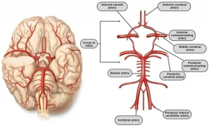
Circulus arteriosus (Circle of Willis)
The cerebral arterial circle (of Willis) is formed at the base of the brain by the interconnecting vertebrobasilar and internal carotid systems of vessels. This anastomotic interconnection is accomplished by:
- An anterior communicating artery connecting the left and right anterior cerebral arteries to each other.
- Two posterior communicating arteries, one on each side, connecting the Internal carotid artery with the posterior cerebral artery.
- The primary purpose of this vascular circle is to provide anastomotic channels if one vessel is occluded.
Physiology of the cerebral cortex, Wernicke’s area, Broca’s area, sensory & motor areas
Cerebrum function, structure, lobes, anatomy, & location
Referred pain examples, Causes of neuropathic pain & Pain control mechanisms
Skull function, anatomy, structure, views & Criteria of neonatal skull
Histological structure of Cerebral cortex & Types of neurons in the cerebral cortex


