Skull function, anatomy, structure, views and Criteria of neonatal skull
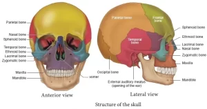
The skull consists of two parts which are the cranium and the mandible, It is a bone structure that forms the head in vertebrates, It supports the structures of the face and provides a protective cavity for the brain, It can protect the brain, It fixes the distance between the eyes to allow stereoscopic vision, and fixes the position of the ears to allow sound localization of the direction and distance of sounds, It is supported by the highest vertebra, called the atlas, permitting nodding motion.
Skull
The skull consists of several separate bones united at immobile joints called sutures, The bones of the skull can be divided into cranial, facial parts, and mandible, It consists of the following bones:
Cranial bones:
- Single: (Fronted, Occipital, Sphenoid, Ethmoid).
- Paired: (Parietal, Temporal).
Facial bones:
- Single: (Vomer, Mandible).
- Paired: (Zygomatic, Maxilla, Nasal, Lacrimal, Palatine, and Inferior concha).
Skull views
The skull will be studied according to the following views (normas):
- Anterior view Norma frontalis)
- Lateral view (Norma lateralis).
- Posterior view (Norma occipitalis).
- Superior view (Norma verticalis).
- Inferior view (Norma basalis) which divides into; norma basalis externa and norma basalis interna.
1. Anterior view (Norma frontalis)
It is formed of the following bones: frontal, nasal, maxilla, zygomatic, and mandible, It shows the following features:
- Supercillary arches, glabella, metopic suture, nasion, supraorbital notch (foramen).
- Orbital margins zygomatico-facial foramen, anterior nasal aperture, anterior nasal spine.
- Infraorbital foramen, alveolar process and alveolar arch.
2. Lateral view (Norma lateralis)
It is formed of the following bones: frontal-parietal, temporal, occipital, zygomatic, and sphenoid, It shows the following features: Coronal suture, lambdoid suture, pterion, superior & inferior temporal lines, temporal fossa, zygomatic arch, zygomatico-temporal foramen external auditory meatus, suprameatal crest, mastoid emissary foramen, mastoid process, and infratemporal fossa.
3. Posterior view (Norma occipitalis)
It is formed of the following bones: parietal temporal, occipital. It shows the following features: sagittal suture, lambdoid suture, lambda, external occipital protuberance, nuchal lines (highest superior and inferior), and external occipital crest.
4. Superior view (Norma verticalis)
It is formed of the following bones: frontal, parietal, and occipital. It shows the following features: coronal suture, sagittal suture, lambdoid suture, bregma, lambda, remnant of metopic suture, parietal emissary foramen, frontal and parietal eminences.
5. Inferior view (Norma basalis externa)
It is formed of the following bones: maxillary, palatine sphenoid, vomer, temporal and occipital bones. It shows the following features:
- Hard palate, incisive fossa & foramen
- Greater & lesser palatine foramina, posterior nasal aperture.
- Medial and lateral pterygoid plates, pterygoid hamulus, pterygoid fossa.
- Scaphoid fossa, mandibular fossa and articular tubercle
- Styloid & mastoid processes, tympanic plate, pharyngeal tubercle.
- Occipital condyles, mastoid notch, and groove for occipital artery.
It has the following important structures:
- Incisive fossa.
- Lesser and greater palatine canals
- Foramina ovale, spinosum, lacerum, jugular, stylomastoid, and foramen magnum
- Canals carotid, hypoglossal, and posterior condylar.
- The bony part of the Eustachian (auditory) tube.
6. Cranial cavity (Norma basalis interna)
Anterior cranial fossa:
- Its floor is formed by the orbital plate of the frontal bone and the cribriform plate of the ethmoid bone.
- In the middle is the crista galli and foramen cecum.
- Lesser wing and anterior part of the body of the sphenoid.
Middle cranial fossa:
Its middle part is formed by the body of sphenoid bone and sphenoidal air sinus. Its lateral part is formed of part of the temporal bone and the petrous part of the temporal bone. It contains many foramina as:
- Optic canal, superior orbital fissure, foramen rotundum.
- Foramen ovale, foramen spinosum and foramen lacerum.
Posterior cranial fossa:
- It is very deep and its floor is formed of the basilar, condylar, and squamous parts of the occipital bone and the mastoid part of the temporal bone.
- It contains many foramina as: Foramen magnum, internal auditory meatus, jugular foramen, and hypoglossal canal.
The foramen & the structure (s) passing through
Foramen cecum: Emissary vein.
Cribriform plate of ethmoid:
- Rootlets of olfactory nerve.
- Anterior & posterior ethmoidal nerve and vessels.
Optic canal:
- Optic nerve.
- Ophthalmic artery
Superior orbital fissure:
- Branches of ophthalmic nerve: (Lacrimal, frontal & nasociliary).
- Oculomotor, trochlear & abducent nerves.
- Ophthalmic veins.
Foramen rotundum:
- Maxillary nerve.
- Middle meningeal vein.
Foramen ovale:
- Mandibular nerve.
- The motor part of the trigeminal nerve.
- Accessory meningeal artery.
- Lesser superficial petrosal nerve.
- Emissary vein.
Foramen spinosum:
- Middle meningeal artery.
- Nervous spinosus.
Foramen lacerum:
- Internal carotid artery & sympathetic plexus around it.
- Greater superficial petrosal nerve.
- Emissary vein.
Internal auditory meatus:
- Facial nerve.
- Vestibulo-cochlear nerve.
- Internal auditory (labrynthine) artery.
Jugular foramen:
- Glossopharyngeal nerve.
- Vagus nerve.
- Accessory nerve.
- Inferior petrosal & sigmoid sinuses.
Hypoglossal canal: Hypoglossal nerve.
Foramen magnum:
- End of medulla & beginning of spinal cord.
- Three layers of meninges.
- Vertebral, ant. & post. spinal arteries.
- Spinal part of the accessory nerve.
- Apical ligament.
Carotid canal: Internal carotid artery.
Lesser palatine canal: Lesser palatine nerve & vessels.
Greater palatine canal: Greater palatine nerve & vessels.
Posterior condylar canal: Emissary vein.
Stylomastoid foramen:
- Facial nerve.
- Stylomastoid artery.
Mandible
The outer surface shows the following features: head, neck mandibular notch, coronoid process, ramus, body angle oblique line, mental foramen, mental tubercle, alveolar process symphysis menti, The inner surface shows the following features: pterygoid fovea, lingula, mandibular foramen, mylohyoid line, and groove, submandibular fossa, sublingual fossa, digastric fossa, mental (genial) spines (2 superior and 2 inferior).
Criteria of neonatal skull
- Proportionately large cranium and small face.
- Smooth, unilaminar, no diploë.
- Fontanelles (anterior [closes at 18th month] and posterior [closes between 6th and 12th month]).
- Ring-shaped tympanic bone.
- The tympanic membrane is nearer to the surface and is directed more inferiorly.
- No mastoid process.
- The mandible is not united (symphysis menti).
- Mandibular angle is obtuse.
Facial muscles function, anatomy, arteries, veins, names and expressions
You can subscribe to science online on YouTube from this link: Science Online
You can download Science Online application on Google Play from this link: Science Online Apps on Google Play


