Electrocardiogram (ECG) importance, ECG test results, analysis and abnormalities
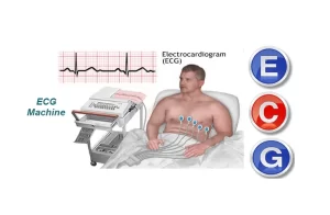
As cardiac impulse passes through the heart, electrical currents spread into tissue surrounding the heart and a small proportion spreads all the way to the surface of the body. If electrodes are placed on the skin on opposite sides of the heart, electrical potentials generated by these currents can be recorded. Each of the cell types in the heart contains a unique electrical discharge pattern (action potential); the sum of these electrical discharges can be recorded as the electrocardiogram (ECG).
The electrocardiogram (ECG)
The electrocardiograph is the instrument, which amplifies and records these electrical potentials on a moving paper strip. The normal ECG is composed of a P wave, a ”QRS complex”, and a T wave. The QRS complex is formed of 3 separate waves, the Q wave, the R wave, and the S wave.
ECG leads
To get a complete picture of the electrical activity of the various parts of the heart, the electrodes are placed at different positions of the body. The recording from these electrodes draws 12 leads on the ECG paper. There are two types of leads: Bipolar leads and unipolar leads.
Bipolar leads
In this type of leads, two active electrodes are used (i.e., bipolar recording), one of them is connected to the negative terminal of the electrocardiograph and the other one to the positive terminal. The electrodes are placed on the right arm, the left arm, and the left leg. There are 3 standard limb leads, which are called, lead I, II, and III, each record the differences in potential between two limbs.
Unipolar leads
Here unipolar recording is obtained by using only one active electrode known as the exploring electrode, which is connected to the positive terminal of the instrument. The second electrode (connected to the negative terminal) is known as the indifferent electrode that stays at zero potential. There are nine unipolar leads: Three unipolar limb leads and six unipolar chest leads.
Unipolar limb leads
Three unipolar limb leads are recorded electrodes, which are placed in the (right arm) VR, (left arm) LV, and (left foot) VF. Augmented limb leads designated by the letter a (aVR, aVL, aVF). The augmented limb leads increase the size of potential by 50% without any change in configuration from the non-augmented record.
The augmented limb leads are unipolar because the potential at the site of the exploring electrodes is compared to zero, not to the potential at another site. The limb leads are arranged to view the heart in the frontal plane (a plane passed vertically through the body as if directly facing it).
- Lead: Position of the exploring electrode.
- a VR: Right arm.
- a VL: Left arm.
- a VF: Left foot.
Unipolar chest
These are six unipolar leads, which the exploring electrodes are arranged on the front wall of the chest, over the heart or precordium. They are designed V1-V6:
- Chest lead: Position of the exploring electrode.
- V1: Right fourth intercostal space near the sternum.
- V2: Left fourth intercostal space near the sternum.
- V3: Midway between V2 and V4.
- V4: Left fifth intercostal space in the midclavicular line.
- V5: Left fifth intercostal space in the anterior axillary line.
- V6: Left fifth intercostal space in the mid-axillary line.
The chest leads are arranged to view the heart in the horizontal (transverse) plane in V1 and V2, the QRS recordings of the normal heart are mainly negative because the chest electrode is nearer the base of the heart than the apex (to the right of the ventricular depolarization wave).
Analysis of the standard ECG
Voltage and time calibration of the ECG: The voltage and duration of the different ECG waves and intervals can be calculated from the calibration lines on the recording ECG paper thus:
- In the vertical direction: (10 small squares) = 1mV.
- In the horizontal direction: 1 small square = 0.04 sec.
- Large square = 5 small squares = 0.2 sec.
Normal ECG
1- The P wave
The P wave is produced by the spread of the depolarization wave from the SA node through the atria to the AV node (from the right to left) positive wave. It represents atrial depolarization. It starts at 0.02 sec. before contraction. The duration of the P wave is 0.1 sec, and its amplitude is 0.1-0.3mV.
Some abnormalities of the P wave
- In atrial hypertrophy the P wave is large.
- In atrial fibrillation, the P wave is replaced by a large number of very fine waves.
2- The QRS complex
The QRS complex is produced by ventricular depolarization. It is composed of three waves.
- The Q wave is the first negative wave, which is produced by depolarization of the inter-ventricular septum, which takes place from left to right, i.e., away from the recording electrode.
- The R wave is the positive wave, which is produced by depolarization of the ventricular muscle from the inner to the outer surface. Since the left ventricle is thicker than the right one, the mean direction of the depolarization wave is downwards and to the left, i.e., towards the recording electrode.
- The S wave is the negative wave following the R wave and is produced by depolarization of the last segment of the myocardium (poster basal portion of the left ventricle and the pulmonary conus), which is directed upwards and to the right i.e., away from the recording electrode.
The QRS complex is known as the initial ventricular contraction. It starts at 0.02 sec. before ventricular contraction. The duration of the QRS complex is 0.04-0.12 sec, and its amplitude is 1-2mV (from top of R to bottom of S).
Some abnormalities of the QRS complex
- Q wave is absent in ventricular septal defects (congenital anomaly). Pathological deep Q wave appears in myocardial infarction.
- The duration of the QRS complex is more than 0.12 sec. in bundle branch block where ventricular depolarization takes a longer time. However, it is shortened when the heart rate is fast as in children and muscular exercise.
- The amplitude of the QRS complex increases in ventricular hypertrophy.
3- The T wave
The T wave is due to the faster and stronger stage of ventricular repolarization. Repolarization of the ventricles starts soon after the end of the S wave but at a very slow rate and does not cause any waves until it becomes strong enough and produces the T wave.
The mean direction of the repolarization wave in the ventricles is opposite to that of the depolarization wave i.e., it spreads away from the recording electrode producing a positive T wave. This is because the ventricular muscle repolarizes from the outer to the inner surface, The duration of the T wave is 0.25 sec, and its amplitude is 0.2-0.3mV.
Some abnormalities of the T wave:
- T wave is flattened in old age.
- T wave is inverted in myocardial ischemia.
Atrial repolarization is not represented in the ECG because it is masked by the QRS complex. Sometimes an additional positive small ”U” wave is present after the T wave, which is believed to be due to slow repolarization of the papillary muscles.
4- The P-R interval
It is the time between the beginnings of the P wave to the beginning of the R wave. Normally it is 0.12-0.2 sec, It represents the atrial depolarization and conduction through the AV node and AV bundle.
Prolongation of the P-R interval occurs if there is impaired conduction from the atria to the ventricles i.e, in the A-V bundle. Thus, the presence of P-R interval exceeding 0.2 sec. indicates the presence of ”first-degree heart block”.
5- The S-T segment
It lies between the end of the S wave and the beginning of the T wave. T wave. It represents the slow ventricular repolarization. It should be isoelectric. Pathologically it may be elevated or depressed.
- The elevation of the S-T segment indicates the acute myocardial injury e.g., due to the recent infarction.
- The depression of the S-T segment occurs during myocardial ischemia, which causes attacks of chest pain known clinically as ”angina pectoris”.
6- The Q-T interval
The Q-T interval is measured from the beginning of the Q wave to the end of the T wave, Its duration is 0.3-0.4 sec, It is a measure of the duration of ventricular contraction. The Q-T interval is shortened in hypercalcaemia and it is prolonged in hypercalcaemia.
Cardiac muscles properties, Conduction system of the heart, Cardiac excitability & contractility
Properties of cardiac muscles, Cardiac automaticity & Conduction of electrical impulses
Histology of the heart, Cardiomyocytes types, Ultrastructure & features of cardiac muscle fibers
Heart & Pericardium structure, Abnormalities & Development of the Heart
Heart function, structure, Valves, Borders, Chambers & Surfaces


