Back muscle anatomy, types, structure, importance and names
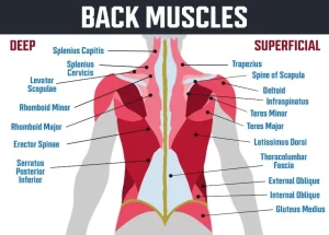
Muscles of the back are divided into superficial, intermediate, and deep muscles, Superficial muscles are associated with movements of the shoulder, Intermediate muscles are associated with movements of the thoracic cage, and Deep muscles are associated with movements of the vertebral column. The deep muscles develop embryologically in the back, and they are described as intrinsic muscles.
Muscles of the back
The superficial layer has Trapezius and Latissimus dorsi. The deep layer has a Levator scapula, Rhomboideus minor, and Rhomboideus major. The superficial back muscles originate from the vertebral column and attach to the shoulder bones – the clavicle, scapula, and humerus.
Trapezius
Origin:
- Superior nuchal line (medial 3/1).
- External occipital protuberance.
- Ligamentum nuchae.
- The spine of the 7th cervical vertebra.
- Supraspinous ligament.
Insertion:
- Clavicle: Posterior border of lateral 1/3rd.
- Scapula: Medial border of acromion, Upper border of the crest of the spine, and Tubercle of the crest of the spine of the scapula.
Nerve supply:
- Motor: Spinal accessory.
- Sensory: From C3, 4 (Proprioception).
Action:
- Upper fibers: Elevation of the shoulder (shrug the shoulder).
- Middle fibers: Retract the scapula.
- Lower fibers: depress the shoulder.
- Upper and lower fibers together rotate the scapula so that the glenoid cavity faces upwards. This occurs when the arm is raised above the head.
Latissimus dorsi muscle
Origin:
- Spinous processes of thoracic T7-T12 and supraspinous ligament.
- Thoracolumbar fascia.
- Outer lip of iliac crest.
- Inferior 3 or 4 ribs.
- The inferior angle of the scapula.
Insertion: Floor of bicipital groove of the humerus.
Nerve supply: Thoracodorsal (nerve to latissimus dorsi) (C6, C7, C8).
Action:
- Adduction, extension, and medially rotation of the arm.
- Climbing (when the arms are fixed above the head).
- Swimming.
- It is a strong muscle of expiration.
Deep layer
The deep layer is formed by the levator scapulae, rhomboideus minor, and major muscles.
Levator scapulae muscle
- Origin: Transverse processes of C1 – C4 vertebrae.
- Insertion in the medial border of the dorsal surface of the scapula from the superior angle to the spine.
- Nerve supply: Dorsal scapular nerve (C5).
- Action: Elevates scapula.
Rhomboideus minor muscle
- Origin is Nuchal ligaments and spinous processes of C7 to T1 vertebrae.
- Insertion: Small area of medial border of the dorsal surface of scapula at the level of the spine, below levator scapulae.
- Nerve supply: Dorsal scapular nerve (C5).
- It retracts the scapula and rotates the scapula so that the glenoid cavity faces downwards.
Rhomboideus major muscle
- Origin is the spinal processes of the T2 to T5 vertebrae.
- Insertion in the Medial border of the scapula, inferior to the insertion of rhomboideus minor muscle.
- The nerve supply is the Dorsal scapular nerve (C5).
- It retracts the scapula and rotates the scapula so that the glenoid cavity faces downwards.
Scapular Region
Supraspinatus muscle
- Origin is supraspinatus fossa (medial 2/3) of scapula.
- Insertion in the Superior facet of greater tuberosity of the humerus.
- The nerve supply is the Suprascapular nerve (C5, C6).
- Action: Abduction of the arm from 0° – 18°.
Infraspinatus muscle
- Origin: Infraspinous fossa (medial 2/3) of the scapula.
- Insertion in the middle facet at the back of the greater tuberosity of the humerus.
- Nerve supply: Suprascapular nerve (C5, C6).
- Action: Lateral rotation of the arm.
Teres minor muscle
- Origin: Lateral border (upper 2/3) of the scapula.
- Insertion in the inferior facet at the back of the greater tuberosity of the humerus.
- Nerve supply: Axillary nerve (C5, C6).
- Action: Lateral rotation and adduction of the arm.
Teres major muscle
- Origin: an oval area on the dorsal surface of the scapula above the inferior angle.
- Insertion: Medical lip of bicipital groove of the humerus.
- Nerve supply: Lower subscapular nerve (C5, C6).
- Action: adduction and medial rotation of the humerus.
Subscapularis muscle
- Origin: Subscapular fossa (medial 2/3).
- Insertion: Lesser tuberosity of the humerus.
- Nerve supply is upper and lower subscapular nerve (C5, C6).
- Action: Adduction and medial rotation of the humerus.
Deltoid muscle
The deltoid muscle is the muscle forming the rounded contour of the shoulder. The middle fibers are multipennate.
Origin:
- Anterior fibers: from the anterior border of lateral 1/3 of the clavicle.
- Middle fibers: from the lateral margin of the acromion.
- Posterior fibers: from the lower lip of the crest of the spine to the scapula.
Insertion: Into the V-shaped deltoid tuberosity in the middle of the lateral aspect of the shaft of the humerus.
Nerve supply: Axillary nerve (C5, C6).
Action:
- Anterior fibers: flexion and medial rotation of the humerus.
- Middle fibers: abduction from 18° – 90°.
- Posterior fibers: extension and lateral rotation of the humerus.
Rotator cuff muscles
Rotator cuff muscles surround the shoulder joint, they include Subscapularis, Supraspinatus, Infraspinatus, and Teres minor.
Intermuscular spaces
Quadrangular space transmits the axillary nerve and posterior circumflex humeral artery.
Boundaries
- Superiorly: Teres minor.
- Inferiorly: Teres major.
- Medially: the long head of the Triceps brachii.
- Laterally: the surgical neck of the humerus.
Medial triangular space contains the circumflex scapular vessels. Boundaries are Teres minor superiorly, Teres major inferiorly, and Long head of the Triceps laterally.
Lateral triangular space contains the Radial nerve and profunda brachii artery.
Boundaries
- Teres major superiorly.
- Long head of the Triceps medially.
- Humerus laterally.
Axillary nerve (Circumflex nerve) (C5,6):
The axillary nerve arises from the posterior cord of the brachial plexus. It passes backwards through the quadrangular space to turn around the surgical neck of the humerus.
Branches:
- Muscular branches: to the deltoid and teres minor muscle.
- Cutaneous branch: Upper lateral cutaneous nerve of the arm which supplies the skin over the lower half of the deltoid.
In the case of a fractured surgical neck humerus, the axillary nerve will be injured and results in weakness of abduction of the arm, wasting of the deltoid muscle (flat shoulder), and loss of sensation over the lower half of the deltoid.
Bones of upper limb structure, function, types and anatomy
Bone (Osseous Tissue) types, structure, function and importance
Bones function, types and structure, The skeleton and Curvature of Spine in Adults
Importance of Anatomical body position, planes and terms of movement
Joints types and function, Nerve supply of joints and general features of Synovial Joints
Types of bones, Histological features of compact bone and cancellous bone


