Upper urinary tract neoplasms symptoms, and What types of tumors are in the urinary tract?
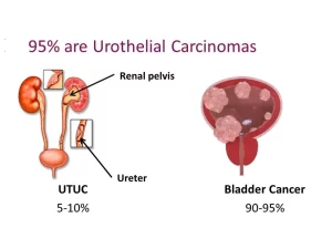
Upper urinary tract neoplasms (UTUC) is a cancer that forms in the lining of the upper urinary tract. This includes the renal pelvis, which is the funnel-shaped area of the kidney where urine collects, and the ureters, which are the two tubes that carry urine from the kidneys to the bladder.
Upper urinary tract neoplasms
UTUC is a relatively uncommon cancer, accounting for about 5% of all urothelial cancers, Urothelial cancers are cancers that start in the cells that line the urinary tract, The most common type of UTUC is urothelial carcinoma, which starts in the transitional cells that line the urinary tract.
Symptoms of UTUs can include:
- Blood in the urine (hematuria).
- Pain in the side or flank.
- A blockage in the urinary tract that can cause pain or difficulty urinating.
- Urinary tract infections (UTIs).
- Weight loss.
- Fatigue.
I- Renal Parenchymal Tumors
Malignant tumors (Renal Cell Carcinoma):
Incidence:
- This is primarily a disease of the elderly patient, with a typical presentation in the sixth and seventh decades of life.
- The majority of cases s of RCC are believed to be sporadic (2% are familial).
- The incidence of RCC has increased due to the more prevalent use of ultrasonography and CT for the evaluation of a variety of abdominal complaints.
Etiology & pathogenesis:
- Tobacco exposure is the most generally accepted environmental risk factor for RCC.
- Obesity is now accepted as another major risk factor for RCC.
- Hypertension appears to be the third major etiologic factor for RCC.
Gross pathology:
- Big tumors bulge outwards from the surface of the kidney.
- Most sporadic RCCs are unilateral and unifocal.
- The cross-section shows a heterogonous appearance: yellow or orange areas (lipids), red areas (hemorrhage), and grey areas (necrosis).
Microscopic examination
1. It is an adenocarcinoma composed of clear cells (lipid and glycogeny granular), granular cells (mitochondria) or both. These cells are arranged in a solid papillary, tubular, or cystic pattern. The prognosis is better with clear-call tumors.
2. There are various histological subtypes;
- The commonest is the clear cell variant.
- Followed by the papillary type.
- Other types include chromophobe cell type and collecting duct type.
3. Clear cell type arises from the Proximal Convoluted Tubules.
Spread
- Hematogenous spread is the earliest line of metastasis because of the rich blood supply of the tumor. The commonest site is the lungs. Other common sites: liver, bone, and brain.
- Lymphatic spread to the para-aortic lymph nodes, and para-caval lymph nodes.
- Direct spread to the perinephric fat and adjacent viscera: colon, pancreas, and adrenal gland.
Clinical Picture
1) Local:
- Because of the sequestered location of the kidney within the retroperitoneum, many renal masses remain asymptomatic and nonpalpable.
- The classic triad of flank pain, gross hematuria, and palpable abdominal mass is now rarely found.
2) Paraneoplastic syndromes (10%-40% of cases): hypertension or (Stauffer syndrome) which is a non-metastatic reversible hepatic dysfunction resulting from metabolic byproducts secreted by the tumor e.g: polycythemia, hypercalcemia.
3) Metastatic: Cough, hemoptysis, dyspnea due to lung metastasis, bone pain-weight loss.
Investigations
- Laboratory: anemia, liver dysfunctions, hypercalcemia.
- Ultrasonography abdomen & pelvis: a minimally invasive tool to detect an echogenic mass in the renal parenchyma. For further assessment by a contrast study.
- Multislice CT scan of abdomen and pelvis (with IV contrast): parenchymal mass with an enhancement after contrast injection. (Diagnostic modality).
Differential Diagnosis: Benign parenchymal tumors e.g. oncocytoma and angiomyolipoma
Treatment:
- Radical nephrectomy: ligation of the renal artery and vein, removal of the whole within the Gerota’s fascia.
- Partial nephrectomy: PN is now the standard of care for the management of clinical T1 renal masses in the presence of a normal contralateral kidney, presuming that the mass is amenable to this approach.
- Systemic therapy: for advanced and metastatic RCC, e.g. targeted therapy and immune therapy.
II. Upper urinary tract urothelial carcinomal
Defined as any neoplastic growth that affects the urothelial lining of the urinary tract from the calyces to the distal ureter.
Incidence
- The peak incidence of upper tract tumors is 10 per 100,000 per year occurring in the age range of 75 to 79 years.
- Accounting for 5% to 7% of all renal tumors and about 5% of all urothelial tumors.
Pathogenesis and Etiology
- Cigarette smoking appears to be the most important of the modifiable risk factors for upper urinary tract urothelial cancer.
- Balkan’s nephropathy (a degenerative interstitial nephropathy). It was first identified among several communities along the Danube River.
- Analgesic abuse is a well-documented risk factor associated with the development of upper urinary tract cancers.
- Chronic Inflammation, Infection. The development of squamous cell cancer is related to chronic bacterial infection associated with urinary stones and obstruction.
Pathology:
Gross appearance:
- Papillary or sessile lesions, and may be unifocal or multifocal.
- Appears in the calyx, renal pelvis, or the whole ureter from the pelviureteric junction to the intramural part of the bladder.
- Ureteral tumors occur more commonly in the lower than in the upper ureter.
Microscopic examination:
- Transitional cell carcinoma: the most common histologic type.
- Squamous cell carcinoma: frequently associated with a condition of chronic inflammation or infection, more likely to be invasive at the time of presentation.
- Adenocarcinoma (very rare).
Clinical Picture
- The most common presenting symptom of upper tract urothelial tumors is hematuria, either gross or microscopic.
- Flank pain is the second most common symptom.
- About 15% of patients are asymptomatic at presentation and are diagnosed when an incidental lesion is found on radiologic evaluation.
- May also present with symptoms of advanced disease, including flank or abdominal mass, weight loss, anorexia, and bone pain.
Investigations
- Multislice CT abdomen and pelvis with IV contrast: the tumor (single or multiple) appears as an irregular filling defect in the lumen of the renal pelvis or ureter. Hydronephrosis is present if the tumor caused an obstructive uropathy.
- Urine cytology: positive for malignant cells in the presence of high-grade tumors as cells shed in the urine (either from the bladder or from the ureter).
- Diagnostic endoscopy: In addition to visualization of the tumor, ureteroscopy allows more accurate biopsy of suspected areas.
Metastasis
- Hematogenous spread is the earliest metastasis because of the rich blood supply of transitional cell carcinoma. The commonest sites of metastasis are the lungs, liver, and bone.
- Lymphatic spread to the para-aortic (renal pelvic and upper ureteric tumors) and pelvic lymph nodes (lower ureteric tumors).
- Direct spread to neighboring tissues (colon, pancreas). A renal pelvic tumor can extend directly to the renal parenchyma.
Treatment
- Radical nephroureterectomy with excision of a bladder cuff is recommended for large, high-grade, invasive tumors of the renal pelvis and proximal ureter.
- Small noninvasive low-grade papillary tumors are ablated with a laser using the ureteroscope (conservative management).
You can subscribe to Science online on YouTube from this link: Science Online
You can download Science Online application on Google Play from this link: Science Online Apps on Google Play
Urinary bladder structure, function, Control of micturition by Brain & Voluntary micturition
Urinary passages function, structure of Ureter, Urinary bladder & Uvulae vesicae
Control of water balance in your body, Regulation of volume & osmolality of Body fluids
Urine formation, Factors affecting Glomerular filtration rate, Tubular reabsorption & secretion
Histological structure of kidneys, Uriniferous tubules & Types of nephrons
Functions of Kidneys, Role of Kidney in glucose homeostasis, Lipid & protein metabolism
Physical properties of urine, Tests to evaluate kidney function & Normal constituents of urine
Packaging of DNA, Genome, chromosomal proteins, DNA in Prokaryotes & Eukaryotes


