Urolithiasis, Stones in the kidney or ureter, How do you treat urolithiasis?
Urinary crystals are microscopic mineral formations that can develop in the urine. They form when there’s an imbalance of certain substances in the urine, such as High levels of minerals, Low urine volume (dehydration), and Abnormal urine pH.
Urinary crystals
Urinary crystals (calcium, phosphate, oxalate, uric acid, xanthine, and cysteine) precipitate and glue together by a (protein matrix of 2-10% of the stone to form the stone. Urinary crystals are usually harmless and don’t cause any symptoms. However, in some cases, they can clump together and form kidney stones, which can be quite painful. There are many different types of urinary crystals, each with its cause.
- Struvite crystals: These crystals are often associated with urinary tract infections (UTIs).
- Calcium oxalate crystals: These are the most common type of urinary crystal and can be caused by dehydration, a diet high in oxalate, or certain medical conditions.
- Uric acid crystals: These crystals can be caused by dehydration, a diet high in protein, or certain medical conditions such as gout.
- Calcium phosphate crystals: These crystals are usually harmless but can sometimes be associated with kidney stones.
- Cystine crystals: These crystals are rare and are caused by a genetic disorder called cystinuria.
Epidemiology
- Gender: adult men are affected with stones 2-3 times more frequently than women.
- Race: in the USA, prevalence is highest among whites and the least among African Americans.
- Age: stone occurrence is relatively (uncommon) before age 20. Age incidence reaches its peak between the 4th and 6th decades of life.
- Climate: a higher prevalence of stone disease is found in hot and dry climates such as deserts and tropical areas (stone belt). This is most pronounced among people who work outdoors. They are exposed to heat and dehydration far more than those who work indoors.
- Water intake: low fluid intake (especially in hot climates) causes dehydration, oliguria, and supersaturation of urine with crystals increasing the risk of stone formation.
Process of Crystallization
The suggested mechanism by which a urinary stone is formed is:
- Supersaturation of urine with crystals is a prerequisite for stone formation.
- Supersaturation initiates crystal nucleation: a small nucleus of precipitated crystals is formed.
- Crystal aggregation and growth occur around the nucleus to form stones of varying sizes.
- Inhibitors of crystal formation e.g. magnesium, citrate, and pyrophosphate reduce the rate of crystal aggregation. A decrease in the level of these inhibitors will enhance stone formation.
Etiology of Urinary Stones
1. Idiopathic: most urinary stones have no known cause for their development.
2. Supersaturation of urine with crystals:
- Hypercalciuria.
- Hyperphosphaturia.
- Hyperoxaluria.
- Hyperuricosuria.
- Cysteinuria.
3. Obstructive uropathy: slowing of the rate of urine flow in the urinary passages or stagnant urine as in a hugely hydronephrotic kidney in congenital pelvi-ureteric junction obstruction, chronic retention bladder or bladder diverticulum, predispose to stone formation.
4. Infection: urinary pathogens, e.g. E coli, can act as a nucleus for aggregation and growth of urinary stones. Also, urea-splitting organisms cause an increase in the urinary pH and precipitation of phosphates.
5. Dehydration: dehydration from reduced fluid intake or massive sweating causes oliguria which in turn, predisposes to supersaturation of urine with crystals and stone formation.
6. Immobilization: chronic immobilization in the presence of a debilitating disease slows the flow of urine, predisposing to stone formation.
Stones in the kidney or ureter
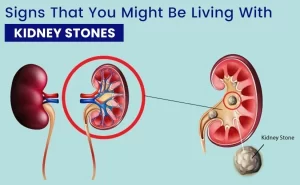
The size of a kidney stone varies from small to very huge and branching stones that can fill the entire pelvicalyccal system (staghorn stones).
Pathogenesis:
- Most urinary stones are formed in the kidney and either remain and grow in the kidney or migrate distally to the ureter.
- The stone will obstruct the kidney or the ureter causing hydronephrosis or hydroureteronephrosis.
- Reflex hyperperistalsis of the urinary tract muscles trying to wash out the stone will cause renal colic and hematuria.
Clinical Presentation
- No symptoms: Most small newly formed stones in a kidney are washed down with urine flow along the ureter and bladder. They are then passed unnoticed along the urethra during micturition.
- Dull aching renal pain: Partial obstruction of the ureter by the stone results in progressive hydroureter and hydronephrosis with subsequent chronic distention of the renal capsule causing persistent mild to moderate chronic dull aching renal pain. The pain is felt in the loin and renal angle and is fixed (no referral)
- Renal colic: A newly formed small stone in the kidney is showered to the ureter with the urine flow. During its descent, it will stimulate the ureteric muscles (at any level) which will undergo sudden reflex spasm around it.
Criteria of renal colic
- Intermittent colicky pain.
- Arise in loin.
- Radiates to the ipsilateral genitalia.
- Associated with nausea and vomiting.
- The patient rolls around, unable to find a comfortable position.
- Maybe associated with low-grade fever.
4. Hematuria: friction from any stone causes bleeding from the urothelium surrounding it. This results in hematuria. In most cases hematuria is microscopic, Gross hematuria can occur if the stone has an irregular surface, especially with spikes protruding from it. Such stones are usually composed of calcium oxalate
5. Lower third ureteric stones cause pain in the urethra and tip of the glans penis with associated irritative lower urinary tract symptoms e.g. urgency and frequency.
6. Fever and general malaise if the stone is impacted and complicated by infected hydronephrosis.
7. Anuria if a stone obstructs a solitary kidney or bilateral obstructing stones.
Diagnosis
1. Non-contrast C.T. abdomen and pelvis (best diagnostic modality): can detect: both radio-opaque and radiolucent stones; and radio-opaque stones that are too small to be seen in a plain X-ray film.
2. Plain X-ray of the abdomen and pelvis (KUB); detects radio-opaque stones (Calcium phosphate, calcium oxalate and calcium phosphate, and cysteine stones). A radio-opaque stone can fail to appear if very small in size. Uric acid stones never appear in plain X-ray films because they are radiolucent.
3. Intravenous urography (IVU) detects radiolucent stones (uric acid) as a filling defect. Other lesions in the urinary tract that cause filling defects are tumors and blood clots.
4. Ultrasonography: can detect stones in the kidney, or bladder. Ultrasound is also useful in showing hydronephrosis if present. It is not useful in the detection of ureteric stones.
5. Laboratory investigations:
- Urine analysis may show crystals, red blood cells, and a few pus cells.
- Blood test for calcium, phosphorus, and uric acid.
- Stone analysis for previously passed stones or removed stones to determine their type.
- Renal functions to exclude uremia.
Treatment
- Emergency treatment.
- Treatment of the stone.
- Preventive measures.
Emergency treatment
- Renal colic: Analgesics (NSAIDs) and antispasmodics.
- Fever due to infected hydronephrosis: intravenous antibiotics and urinary diversion by PCN or DJ.
- Anuria: Urinary diversion by PCN or DJ. Hemodialysis in case of uncompensated uremia.
Treatment of the stone
- Lines of treatment.
- Treatment according to stone location and stone size.
Lines of treatment
1. Medical treatment: indicated in small stones less than 0.5cm.
- Analgesics and antispasmodics.
- Diuresis by increasing fluid intake.
- Alpha-blockers for lower ureteric stones induce ureteric muscle relaxation.
- Periodic radiologic examination for follow-up.
- For radiolucent stones (uric acid stones): Medical alkalization of the urine by potassium citrate and sodium bicarbonate for stone dissolution.
2. Extracorporeal shock wave lithotripsy (ESWL).
i. Indications:
- Kidney stones ≤ 2cm.
- Proximal ureteric stones ≤ 1cm.
ii. Contraindications:
- Uncorrectable bleeding disorders or patients on anticoagulants.
- Pregnancy.
- Obstruction distal to the stone.
iii. Complications:
- Ureteral obstruction by stone fragments (steinstrasse).
- Hematuria.
- Subcapsular bleeding and peri-renal hematoma.
3. Endoscopic treatment
a) Percutaneous nephrolithotomy (PCNL): indicated in kidney stones > 2cm. This procedure entails:
- Percutaneous puncture of the kidney by a special needle under fluoroscopy where the needle passes from the skin to the collecting system.
- Dilatation of the tract by special dilators.
- Introduction of the nephroscope through the dilated tract.
- Pneumatic lithotripsy of the stone into small fragments.
- Extraction of the fragments by forceps.
b) Flexible ureteroscopy and laser lithotripsy for stones in the upper third ureter > 1cm and in kidney stones as alternative to ESWL (When ESWL is contraindicated as in pregnancy and coagulopathy).
c) Semirigid ureteroscopy and laser lithotripsy for distal ureteric stones not responding to medical treatment.
4. Surgical treatment
- Pyelolithotomy, and nephrolithotomy for large kidney stones.
- Ureterolithotomy for large ureteric stones.
- Nephrectomy for nonfunctioning kidney.
Treatment according to stone location and stone size
A. Kidney stones
- Small stones less than 0.5cm: Expectant medical treatment.
- Stones ≤ 2cm: ESWL.
- Stones > 2cm: PCNL.
- Staghorn stone: Surgical treatment or PCNL + ESWL.
- Stones with nonfunctioning kidney: Nephrectomy.
B. Proximal ureteric stones:
- Small stones less than 0.5 cm: Expectant medical treatment.
- Stones ≤ 1cm: ESWL.
- Stones 1cm to 2cm: Flexible ureteroscopy.
- Large stones: Surgical treatment (ureterolithotomy).
C. Middle ureteric stones:
- Small stones less than 0.5cm: Expectant medical treatment.
- Stones up to 2cm: Ureteroscopy (flexible semirigid) and laser lithotripsy.
- Large stones: Surgical treatment) (ureterolithotomy).
D. Lower third ureteric stones:
- Small stones less than 0.5cm: Expectant medical treatment.
- Stones up to 2cm: Semirigid ureteroscopy and laser lithotripsy.
- Large stones: Surgical treatment (ureterolithotomy).
III. Preventive measures:
- Balanced diet.
- Increase fluid intake.
- Adequate treatment of UTI or obstruction.
- Treatment of general factors such as hyperthyroidism and gout.
You can subscribe to Science Online on YouTube from this link: Science Online
You can download Science Online application on Google Play from this link: Science Online Apps on Google Play
Anomalies of the bladder and urethra, Urinary tract abnormalities symptoms
What are urinary tract infections?, kidney problems, Urinary retention and Bladder stones
Functions of Kidneys, Role of Kidney in glucose homeostasis, Lipid & protein metabolism
Histological structure of kidneys, Uriniferous tubules and Types of nephrons
Urine formation, Factors affecting Glomerular filtration rate, Tubular reabsorption and secretion
Urinary passages function, structure of Ureter, Urinary bladder & Uvulae vesicae
Urinary system structure, function, anatomy, organs, Blood supply and Importance of renal fascia
Urinary bladder structure, function, Control of micturition by Brain & Voluntary micturition


